Canary Institute for Cancer Research (ICIC), Las Palmas, Spain.
Radiat Oncol. 2010 Jan 28;5:4. doi: 10.1186/1748-717X-5-4.
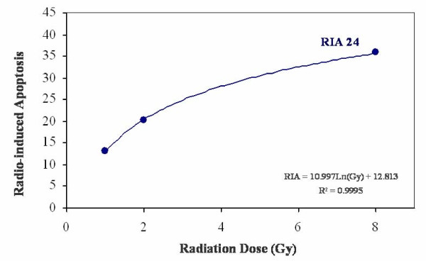
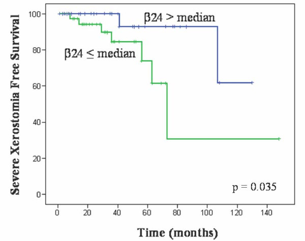
Head and neck cancer is treated mainly by surgery and radiotherapy. Normal tissue toxicity due to x-ray exposure is a limiting factor for treatment success. Many efforts have been employed to develop predictive tests applied to clinical practice. Determination of lymphocyte radio-sensitivity by radio-induced apoptosis arises as a possible method to predict tissue toxicity due to radiotherapy. The aim of the present study was to analyze radio-induced apoptosis of peripheral blood lymphocytes in head and neck cancer patients and to explore their role in predicting radiation induced toxicity. Seventy nine consecutive patients suffering from head and neck cancer, diagnosed and treated in our institution, were included in the study. Toxicity was evaluated using the Radiation Therapy Oncology Group scale. Peripheral blood lymphocytes were isolated and irradiated at 0, 1, 2 and 8 Gy during 24 hours. Apoptosis was measured by flow cytometry using annexin V/propidium iodide. Lymphocytes were marked with CD45 APC-conjugated monoclonal antibody. Radiation-induced apoptosis increased in order to radiation dose and fitted to a semi logarithmic model defined by two constants: alpha and beta. Alpha, as the origin of the curve in the Y axis determining the percentage of spontaneous cell death, and beta, as the slope of the curve determining the percentage of cell death induced at a determined radiation dose, were obtained. beta value was statistically associated to normal tissue toxicity in terms of severe xerostomia, as higher levels of apoptosis were observed in patients with low toxicity (p = 0.035; Exp(B) 0.224, I.C.95% (0.060-0.904)). These data agree with our previous results and suggest that it is possible to estimate the radiosensitivity of peripheral blood lymphocytes from patients determining the radiation induced apoptosis with annexin V/propidium iodide staining. beta values observed define an individual radiosensitivity profile that could predict late toxicity due to radiotherapy in locally advanced head and neck cancer patients. Anyhow, prospective studies with different cancer types and higher number of patients are needed to validate these results.
头颈部癌症主要通过手术和放疗进行治疗。由于 X 射线照射导致的正常组织毒性是治疗成功的限制因素。为了将预测测试应用于临床实践,已经做出了许多努力。通过放射诱导的细胞凋亡来确定淋巴细胞的放射敏感性,这是一种预测放疗引起的组织毒性的可能方法。本研究的目的是分析头颈部癌症患者外周血淋巴细胞的放射诱导凋亡,并探讨其在预测放射诱导毒性中的作用。纳入了在我们机构诊断和治疗的 79 例连续的头颈部癌症患者。使用放射治疗肿瘤学组(Radiation Therapy Oncology Group)量表评估毒性。在 24 小时内,将外周血淋巴细胞分离并在 0、1、2 和 8 Gy 下照射。通过流式细胞术使用 Annexin V/碘化丙啶测量凋亡。用 CD45 APC 缀合的单克隆抗体标记淋巴细胞。放射诱导的凋亡随放射剂量增加而增加,并拟合到由两个常数定义的半对数模型:α和β。α作为 Y 轴上曲线的原点,用于确定自发细胞死亡的百分比,β作为曲线的斜率,用于确定在特定放射剂量下诱导的细胞死亡百分比。β值在严重口干症的正常组织毒性方面与统计学相关,因为低毒性患者的凋亡水平更高(p=0.035;Exp(B)0.224,95%置信区间(0.060-0.904))。这些数据与我们之前的结果一致,并表明通过用 Annexin V/碘化丙啶染色确定放射诱导的凋亡,可以从患者中估计外周血淋巴细胞的放射敏感性。观察到的β值定义了一个个体放射敏感性谱,可以预测局部晚期头颈部癌症患者放疗后的迟发性毒性。无论如何,需要进行具有不同癌症类型和更多患者的前瞻性研究来验证这些结果。