Spinal Cord and Brain Injury Research Center, Chandler Medical Center, University of Kentucky, USA.
Brain Res. 2010 Apr 6;1323:161-73. doi: 10.1016/j.brainres.2010.01.067. Epub 2010 Feb 1.
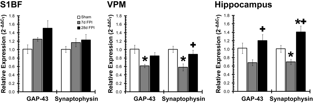
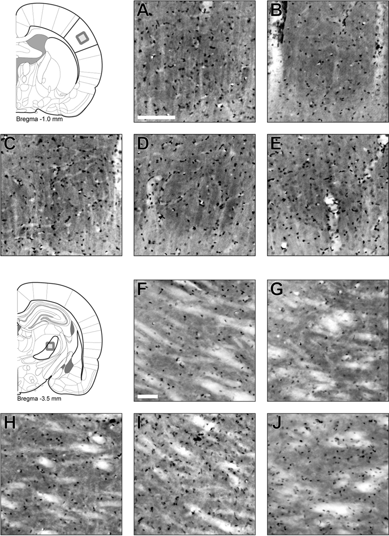
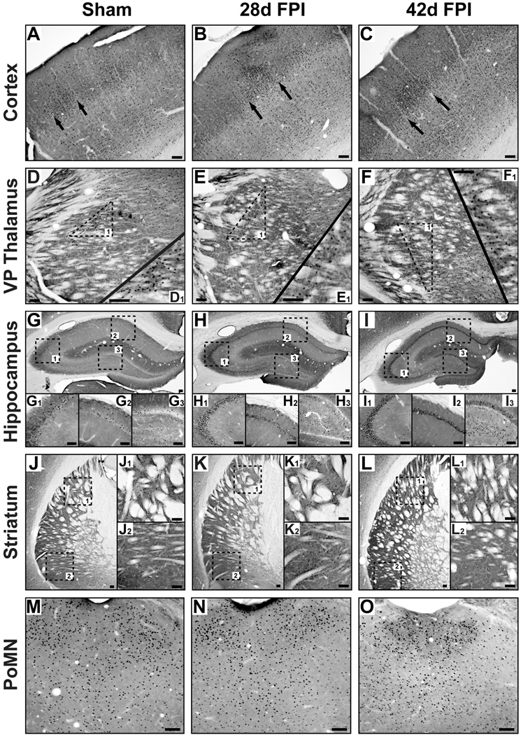
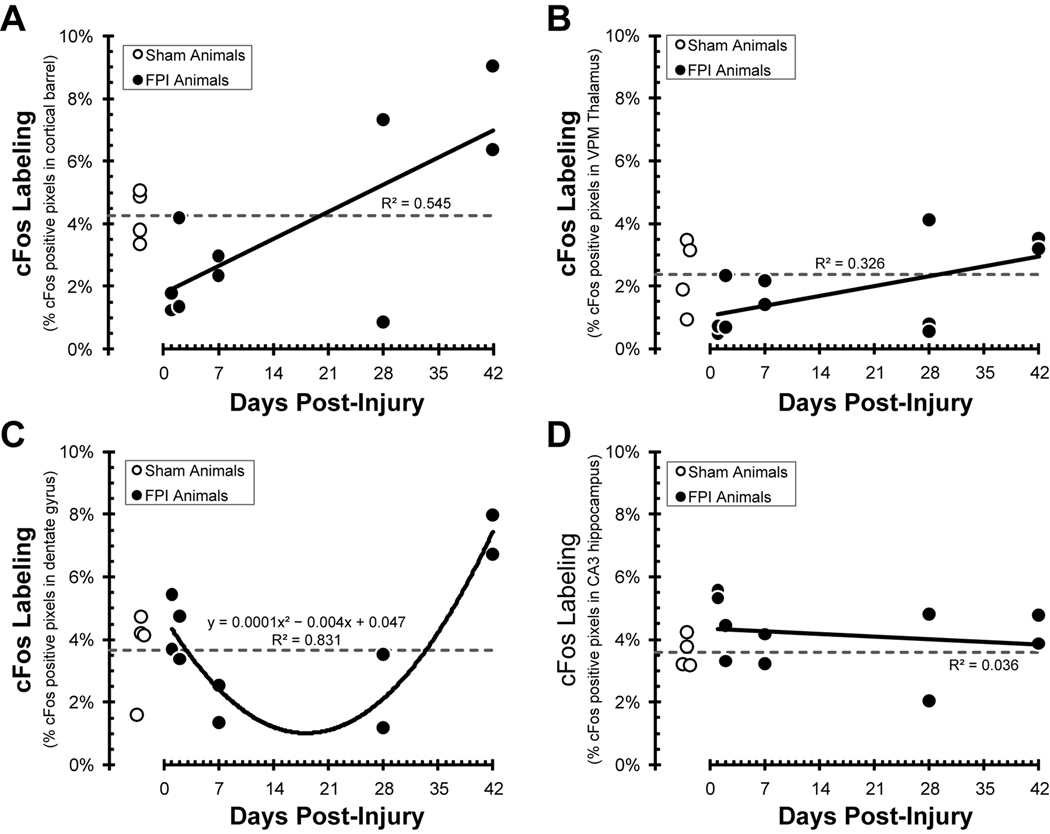
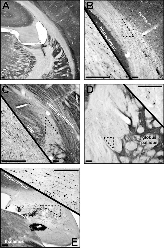
Traumatic brain injury can initiate an array of chronic neurological deficits, effecting executive function, language and sensorimotor integration. Mechanical forces produce the diffuse pathology that disrupts neural circuit activation across vulnerable brain regions. The present manuscript explores the hypothesis that the extent of functional activation of brain-injured circuits is a consequence of initial disruption and consequent reorganization. In the rat, enduring sensory sensitivity to whisker stimulation directs regional analysis to the whisker barrel circuit. Adult, male rats were subjected to midline fluid percussion brain or sham injury and evaluated between 1day and 42days post-injury. Whisker somatosensory regions of the cortex and thalamus maintained cellular composition as visualized by Nissl stain. Within the first week post-injury, quantitatively less cFos activation was elicited by whisker stimulation, potentially due to axotomy within and surrounding the whisker circuit as visualized by amyloid precursor protein immunohistochemistry. Over six weeks post-injury, cFos activation after whisker stimulation showed a significant linear correlation with time in the cortex (r(2)=0.545; p=0.015), non-significant correlation in the thalamus (r(2)=0.326) and U-shaped correlation in the dentate gyrus (r(2)=0.831), all eventually exceeding sham levels. Ongoing neuroplastic responses in the cortex are evidenced by accumulating growth associated protein and synaptophysin gene expression. In the thalamus, the delayed restoration of plasticity markers may explain the broad distribution of neuronal activation extending into the striatum and hippocampus with whisker stimulation. The sprouting of diffuse-injured circuits into diffuse-injured tissue likely establishes maladaptive circuits responsible for behavioral morbidity. Therapeutic interventions to promote adaptive circuit restructuring may mitigate post-traumatic morbidity.
创伤性脑损伤会引发一系列慢性神经功能缺陷,影响执行功能、语言和感觉运动整合。机械力产生弥漫性病变,破坏易损脑区的神经回路激活。本文探讨了这样一种假设,即脑损伤回路的功能激活程度是初始破坏和随后重组的结果。在大鼠中,对胡须刺激的持久感觉敏感性将区域分析指向胡须桶状回路。成年雄性大鼠接受中线液冲击性脑损伤或假手术,并在损伤后 1 天至 42 天进行评估。皮质和丘脑的胡须体感区保持细胞组成,如尼氏染色所示。在损伤后第一周内,由胡须刺激引起的 cFos 激活定量减少,可能是由于胡须回路内和周围的轴突切断,如淀粉样前体蛋白免疫组化所示。在损伤后六周内,由胡须刺激引起的 cFos 激活与皮质中的时间呈显著线性相关(r²=0.545;p=0.015),在丘脑呈非显著相关(r²=0.326),在齿状回呈 U 形相关(r²=0.831),所有这些最终都超过了假手术水平。皮质中持续的神经可塑性反应表现为生长相关蛋白和突触小体蛋白基因表达的积累。在丘脑,可塑性标志物的延迟恢复可能解释了随着胡须刺激,神经元激活向纹状体和海马区广泛分布的现象。弥漫性损伤回路向弥漫性损伤组织的发芽可能建立了导致行为发病率的适应性不良回路。促进适应性回路重构的治疗干预可能减轻创伤后发病率。