• 文献检索
  • 文档翻译
  • 深度研究
  • 学术资讯
  • Suppr Zotero 插件Zotero 插件
  • 邀请有礼
  • 套餐&价格
  • 历史记录
应用&插件
Suppr Zotero 插件Zotero 插件浏览器插件Mac 客户端Windows 客户端微信小程序
定价
高级版会员购买积分包购买API积分包
服务
文献检索文档翻译深度研究API 文档MCP 服务
关于我们
关于 Suppr公司介绍联系我们用户协议隐私条款
关注我们

Suppr 超能文献

核心技术专利:CN118964589B侵权必究
粤ICP备2023148730 号-1Suppr @ 2026

文献检索

告别复杂PubMed语法,用中文像聊天一样搜索,搜遍4000万医学文献。AI智能推荐,让科研检索更轻松。

立即免费搜索

文件翻译

保留排版,准确专业,支持PDF/Word/PPT等文件格式,支持 12+语言互译。

免费翻译文档

深度研究

AI帮你快速写综述,25分钟生成高质量综述,智能提取关键信息,辅助科研写作。

立即免费体验

磁共振成像在成人眼眶良恶性肿瘤鉴别诊断中的价值。

Value of MR imaging in the differentiation of benign and malignant orbital tumors in adults.

机构信息

Department of Radiology, Capital Medical University, Beijing Tongren Hospital, Beijing, China.

出版信息

Eur Radiol. 2010 Jul;20(7):1692-702. doi: 10.1007/s00330-009-1711-0. Epub 2010 Feb 4.

DOI:10.1007/s00330-009-1711-0
PMID:20131055
原文链接:https://pmc.ncbi.nlm.nih.gov/articles/PMC2882044/
Abstract

PURPOSE

To prospectively evaluate magnetic resonance (MR) imaging including dynamic contrast-enhanced MR imaging in the differentiation of benign from malignant orbital masses and to evaluate which MR imaging features are most predictive of malignant tumors.

MATERIALS AND METHODS

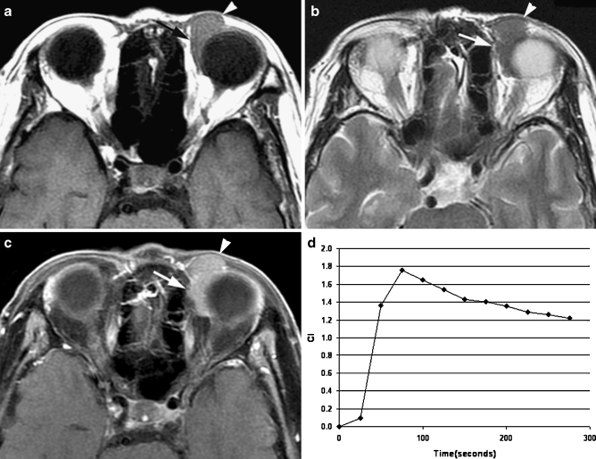
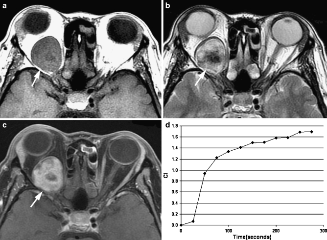
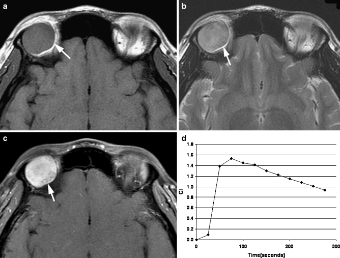
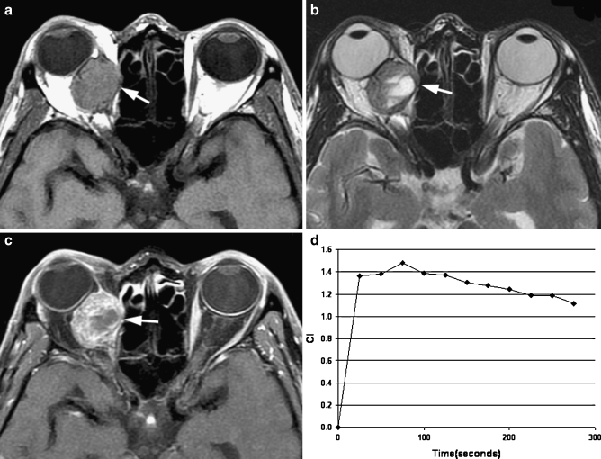
The study was approved by the institutional review board and signed informed consent was obtained. Nonenhanced, static, and dynamic contrast-enhanced MR imaging was performed in 102 adult patients with an orbital mass. Diagnosis was based on histologic findings. MR imaging features of benign and malignant orbital lesions were evaluated correlated with histological findings. Multivariate logistic regression analysis was employed to identify the best combination of MR imaging features that might be predictive of malignancy.

RESULTS

Nonenhanced, static, and dynamic enhancement MR imaging was significantly superior to two other models in prediction of malignancy (p < 0.05). Multivariate logistic regression analysis identified that the most discriminating MR imaging features were isointense mass on T2-weighted imaging and a washout-type time-intensity curve for both observers.

CONCLUSION

Nonenhanced, static, and dynamic enhancement MR imaging improved differentiation between benign and malignant orbital masses in adult patients.

摘要

目的

前瞻性评估磁共振成像(MR 成像),包括动态对比增强 MR 成像,以区分良性和恶性眼眶肿块,并评估哪些 MR 成像特征对恶性肿瘤最具预测性。

材料与方法

本研究经机构审查委员会批准,并获得患者签署的知情同意书。102 例成人眼眶肿块患者进行了非增强、静态和动态对比增强 MR 成像。诊断基于组织学发现。评估了良性和恶性眼眶病变的 MR 成像特征,并与组织学发现相关联。采用多变量逻辑回归分析确定最能预测恶性肿瘤的最佳 MR 成像特征组合。

结果

非增强、静态和动态增强 MR 成像在预测恶性肿瘤方面明显优于其他两种模型(p<0.05)。多变量逻辑回归分析确定最具鉴别力的 MR 成像特征是 T2 加权成像上等信号肿块和两位观察者的洗脱型时间-强度曲线。

结论

非增强、静态和动态增强 MR 成像改善了成人眼眶良恶性肿块的鉴别诊断。

https://cdn.ncbi.nlm.nih.gov/pmc/blobs/8c42/2882044/575071c031ae/330_2009_1711_Fig4_HTML.jpg
https://cdn.ncbi.nlm.nih.gov/pmc/blobs/8c42/2882044/6ba948806420/330_2009_1711_Fig1_HTML.jpg
https://cdn.ncbi.nlm.nih.gov/pmc/blobs/8c42/2882044/ddb2dd8c9a12/330_2009_1711_Fig2_HTML.jpg
https://cdn.ncbi.nlm.nih.gov/pmc/blobs/8c42/2882044/f5c19fd4e382/330_2009_1711_Fig3_HTML.jpg
https://cdn.ncbi.nlm.nih.gov/pmc/blobs/8c42/2882044/575071c031ae/330_2009_1711_Fig4_HTML.jpg

相似文献

1
Value of MR imaging in the differentiation of benign and malignant orbital tumors in adults.磁共振成像在成人眼眶良恶性肿瘤鉴别诊断中的价值。
Eur Radiol. 2010 Jul;20(7):1692-702. doi: 10.1007/s00330-009-1711-0. Epub 2010 Feb 4.
2
Soft-tissue tumors: value of static and dynamic gadopentetate dimeglumine-enhanced MR imaging in prediction of malignancy.软组织肿瘤:钆喷酸葡胺增强磁共振成像的静态和动态表现对预测肿瘤恶性程度的价值
Radiology. 2004 Nov;233(2):493-502. doi: 10.1148/radiol.2332031110. Epub 2004 Sep 30.
3
Assessment of dynamic contrast-enhanced magnetic resonance imaging in the differentiation of malignant from benign orbital masses.动态对比增强磁共振成像在眼眶良恶性肿块鉴别中的评估。
Eur J Radiol. 2013 Sep;82(9):1506-11. doi: 10.1016/j.ejrad.2013.03.001. Epub 2013 Apr 3.
4
Differentiation of malignant from benign pheochromocytomas with diffusion-weighted and dynamic contrast-enhanced magnetic resonance at 3.0 T.3.0T磁共振扩散加权成像与动态对比增强成像鉴别恶性与良性嗜铬细胞瘤
J Comput Assist Tomogr. 2012 Jul-Aug;36(4):361-6. doi: 10.1097/RCT.0b013e31825975f8.
5
Orbital Indeterminate Lesions in Adults: Combined Magnetic Resonance Morphometry and Histogram Analysis of Apparent Diffusion Coefficient Maps for Predicting Malignancy.成人眼眶不确定性病变:联合磁共振形态测量和表观扩散系数图的直方图分析预测恶性肿瘤
Acad Radiol. 2016 Feb;23(2):200-8. doi: 10.1016/j.acra.2015.10.015. Epub 2015 Nov 28.
6
Diffusion-weighted imaging improves the diagnostic accuracy of conventional 3.0-T breast MR imaging.弥散加权成像提高了常规 3.0-T 乳腺磁共振成像的诊断准确性。
Radiology. 2010 Jul;256(1):64-73. doi: 10.1148/radiol.10091367.
7
Dynamic bilateral contrast-enhanced MR imaging of the breast: trade-off between spatial and temporal resolution.乳腺动态双侧对比增强磁共振成像:空间分辨率与时间分辨率之间的权衡
Radiology. 2005 Sep;236(3):789-800. doi: 10.1148/radiol.2363040811.
8
Characterization of orbital masses by multiparametric MRI.通过多参数磁共振成像对眼眶肿物进行特征描述。
Eur J Radiol. 2016 Feb;85(2):324-36. doi: 10.1016/j.ejrad.2015.11.041. Epub 2015 Dec 4.
9
CT-MR image data fusion for computer-assisted navigated surgery of orbital tumors.CT-MR 图像数据融合在计算机辅助眼眶肿瘤导航手术中的应用。
Eur J Radiol. 2010 Feb;73(2):224-9. doi: 10.1016/j.ejrad.2008.11.003. Epub 2008 Dec 20.
10
The value of contrast-enhanced dynamic and diffusion-weighted MR imaging for distinguishing benign and malignant splenic masses.对比增强动态磁共振成像和扩散加权磁共振成像在鉴别脾脏良恶性肿块中的价值。
Br J Radiol. 2016 Jul;89(1063):20160054. doi: 10.1259/bjr.20160054. Epub 2016 May 10.

引用本文的文献

1
Deep learning-based intratumoral and peritumoral features for differentiating ocular adnexal lymphoma and idiopathic orbital inflammation.基于深度学习的肿瘤内和肿瘤周围特征用于鉴别眼附属器淋巴瘤和特发性眼眶炎症。
Eur Radiol. 2025 Mar;35(3):1276-1289. doi: 10.1007/s00330-024-11275-5. Epub 2024 Dec 19.
2
Diagnostic Performance of Dynamic Contrast-Enhanced 3T MR Imaging for Characterization of Orbital Lesions: Validation in a Large Prospective Study.动态对比增强3T磁共振成像对眼眶病变特征的诊断效能:一项大型前瞻性研究中的验证
AJNR Am J Neuroradiol. 2024 Mar 7;45(3):342-350. doi: 10.3174/ajnr.A8131.
3
End-to-End Deep-Learning-Based Diagnosis of Benign and Malignant Orbital Tumors on Computed Tomography Images.

本文引用的文献

1
Orbital lymphoproliferative tumors: analysis of clinical features and systemic involvement in 160 cases.眼眶淋巴增生性肿瘤:160例临床特征及全身受累情况分析
Ophthalmology. 2008 Sep;115(9):1626-31, 1631.e1-3. doi: 10.1016/j.ophtha.2008.02.004. Epub 2008 Apr 28.
2
Imaging features of ocular adnexal lymphoproliferative disease.眼附属器淋巴增殖性疾病的影像学特征。
Eye (Lond). 2006 Oct;20(10):1189-95. doi: 10.1038/sj.eye.6702382.
3
Magnetic resonance imaging of orbital tumors.眼眶肿瘤的磁共振成像
基于深度学习的计算机断层扫描图像上眼眶良恶性肿瘤的端到端诊断
J Pers Med. 2023 Jan 23;13(2):204. doi: 10.3390/jpm13020204.
4
Sinonasal lymphoma: A primer for otolaryngologists.鼻窦淋巴瘤:耳鼻喉科医生入门指南。
Laryngoscope Investig Otolaryngol. 2022 Oct 4;7(6):1712-1724. doi: 10.1002/lio2.941. eCollection 2022 Dec.
5
A signature of structural MRI features at 3 Tesla allows an accurate characterization of orbital cavernous venous malformation.3特斯拉下结构磁共振成像特征的一个标志能够准确表征眼眶海绵状静脉畸形。
Eur Radiol. 2023 Mar;33(3):2149-2159. doi: 10.1007/s00330-022-09163-x. Epub 2022 Oct 20.
6
Clinical and imaging clues to the diagnosis and follow-up of ptosis and ophthalmoparesis.眼睑下垂和眼肌瘫痪的诊断和随访的临床及影像学线索。
J Cachexia Sarcopenia Muscle. 2022 Dec;13(6):2820-2834. doi: 10.1002/jcsm.13089. Epub 2022 Sep 29.
7
A deep learning model combining multimodal radiomics, clinical and imaging features for differentiating ocular adnexal lymphoma from idiopathic orbital inflammation.一种结合多模态放射组学、临床和影像学特征的深度学习模型,用于鉴别眼附属器淋巴瘤与特发性眼眶炎症。
Eur Radiol. 2022 Oct;32(10):6922-6932. doi: 10.1007/s00330-022-08857-6. Epub 2022 Jun 8.
8
MRI-Based Radiomics for Differentiating Orbital Cavernous Hemangioma and Orbital Schwannoma.基于磁共振成像的影像组学用于鉴别眼眶海绵状血管瘤和眼眶神经鞘瘤
Front Med (Lausanne). 2021 Dec 16;8:795038. doi: 10.3389/fmed.2021.795038. eCollection 2021.
9
Imaging of head and neck mucosa-associated lymphoid tissue lymphoma (MALToma).头颈部黏膜相关淋巴组织淋巴瘤(MALToma)的影像学表现。
Cancer Imaging. 2021 Jan 12;21(1):10. doi: 10.1186/s40644-020-00380-5.
10
Quantitative characterization of extraocular orbital lesions in children using diffusion-weighted imaging.应用扩散加权成像对儿童眼眶外病变的定量特征分析。
Pediatr Radiol. 2021 Jan;51(1):119-127. doi: 10.1007/s00247-020-04807-w. Epub 2020 Sep 8.
Eur Radiol. 2006 Oct;16(10):2207-19. doi: 10.1007/s00330-006-0227-0. Epub 2006 Apr 1.
4
Diagnostic architectural and dynamic features at breast MR imaging: multicenter study.乳腺磁共振成像的诊断性结构和动态特征:多中心研究
Radiology. 2006 Jan;238(1):42-53. doi: 10.1148/radiol.2381042117.
5
Rethinking orbital imaging establishing guidelines for interpreting orbital imaging studies and evaluating their predictive value in patients with orbital tumors.重新审视眼眶成像:制定眼眶成像研究解读指南并评估其在眼眶肿瘤患者中的预测价值。
Ophthalmology. 2005 Dec;112(12):2196-207. doi: 10.1016/j.ophtha.2005.09.013.
6
Orbital schwannoma and neurofibroma: role of imaging.眼眶神经鞘瘤和神经纤维瘤:影像学的作用
Neuroimaging Clin N Am. 2005 Feb;15(1):159-74. doi: 10.1016/j.nic.2005.02.004.
7
Orbital cavernous hemangioma: role of imaging.眼眶海绵状血管瘤:影像学的作用
Neuroimaging Clin N Am. 2005 Feb;15(1):137-58. doi: 10.1016/j.nic.2005.02.009.
8
Orbital imaging: Part 2. Intraorbital pathology.眼眶成像:第2部分。眶内病理学
Clin Radiol. 2005 Mar;60(3):288-307. doi: 10.1016/j.crad.2004.05.018.
9
Differentiation of cavernous hemangioma from schwannoma of the orbit: a dynamic MRI study.眼眶海绵状血管瘤与神经鞘瘤的鉴别:动态MRI研究
AJR Am J Roentgenol. 2004 Dec;183(6):1799-804. doi: 10.2214/ajr.183.6.01831799.
10
Soft-tissue tumors: value of static and dynamic gadopentetate dimeglumine-enhanced MR imaging in prediction of malignancy.软组织肿瘤:钆喷酸葡胺增强磁共振成像的静态和动态表现对预测肿瘤恶性程度的价值
Radiology. 2004 Nov;233(2):493-502. doi: 10.1148/radiol.2332031110. Epub 2004 Sep 30.