Suppr超能文献

衰减全反射红外显微光谱成像方法在肾活检分析中的优势。

The advantages of an attenuated total internal reflection infrared microspectroscopic imaging approach for kidney biopsy analysis.

机构信息

Molecular Microspectroscopy Laboratory, Department of Chemistry and Biochemistry, Miami University, Oxford, Ohio 45056, USA.

出版信息

Appl Spectrosc. 2010 Jan;64(1):15-22. doi: 10.1366/000370210792966161.

Abstract

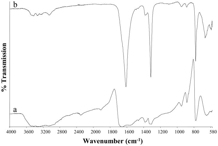
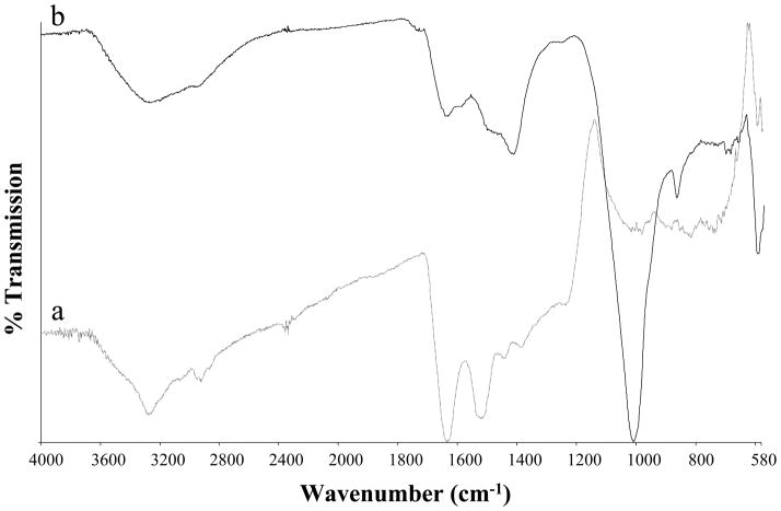
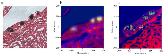
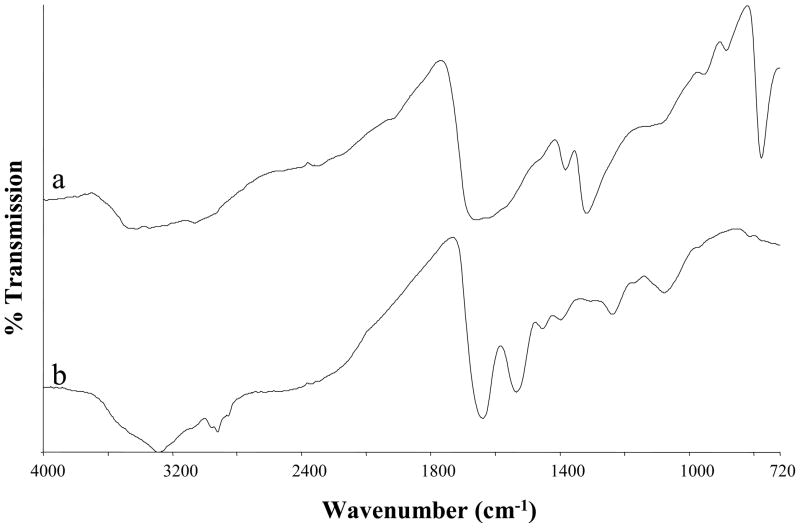
The benefits of an attenuated total reflection Fourier transform infrared (ATR-FTIR) imaging approach for kidney biopsy analysis are described. Biopsy sections collected from kidney-stone formers are analyzed at the initial stages of stone development to provide insights into stone growth and formation. The majority of tissue analysis currently conducted with IR microspectroscopy is performed with a transflection method. The research presented in this manuscript demonstrates that ATR overcomes many of the disadvantages of transflection or transmission measurements for tissue analysis including an elimination of spectral artifacts. When kidney biopsies with small mineral inclusions are analyzed with a transflection approach, specular reflection and the Christiansen effect (anomalous dispersion) can occur, leading to spectral artifacts. Another effect specific to the analysis of mineral inclusions present in kidney biopsies is known as the reststrahlen effect whereby the inclusions become strong reflectors near an absorption band. ATR eliminates these effects by immersing the sample in a high index medium. Additionally, the focused beam size for ATR is decreased by a factor of four when a germanium internal reflection element is used, allowing the acquisition of spectra from small mineral inclusions several micrometers in diameter. If quantitative analysis of small mineral inclusions is ultimately desired, ATR provides the photometrically accurate spectra necessary for quantification.

摘要

本文介绍了衰减全反射傅里叶变换红外(ATR-FTIR)成像方法在肾活检分析中的应用。从结石形成者的肾活检组织中采集样本,在结石形成的初始阶段进行分析,以深入了解结石的生长和形成过程。目前,大多数采用红外光谱显微镜进行的组织分析都是采用反射法进行的。本文介绍的研究表明,ATR 克服了反射或透射测量在组织分析中存在的许多缺点,包括消除光谱伪影。当采用反射法分析含有小矿物包裹体的肾活检组织时,可能会出现镜面反射和克里斯琴森效应(异常色散),导致光谱伪影。另一种特定于分析肾活检组织中矿物包裹体的效应称为瑞利后向散射效应,即包裹体在吸收带附近成为强反射体。ATR 通过将样品浸入高折射率介质中消除了这些效应。此外,当使用锗内反射元件时,ATR 的聚焦光束尺寸减小了四倍,允许从直径几微米的小矿物包裹体中获取光谱。如果最终需要对小矿物包裹体进行定量分析,ATR 则提供了进行定量所需的光度准确的光谱。

相似文献

2
A concerted protocol for the analysis of mineral deposits in biopsied tissue using infrared microanalysis.
Urol Res. 2005 Jun;33(3):213-9. doi: 10.1007/s00240-004-0456-0. Epub 2005 Feb 10.
7
Attenuated total reflection Fourier transform infrared (ATR-FTIR) spectroscopy of a single endothelial cell.
Analyst. 2012 Sep 21;137(18):4135-9. doi: 10.1039/c2an35331h. Epub 2012 Aug 2.
10
Analysis of urinary calculi using an infrared microspectroscopic surface reflectance imaging technique.
Urol Res. 2007 Feb;35(1):41-8. doi: 10.1007/s00240-006-0077-x. Epub 2007 Jan 5.

引用本文的文献

1
Sample preparation and analysis protocols for the elucidation of structure and chemical distribution in kidney stones.
Spectrochim Acta A Mol Biomol Spectrosc. 2024 Jan 15;305:123561. doi: 10.1016/j.saa.2023.123561. Epub 2023 Oct 18.
2
The Convergence of FTIR and EVs: Emergence Strategy for Non-Invasive Cancer Markers Discovery.
Diagnostics (Basel). 2022 Dec 21;13(1):22. doi: 10.3390/diagnostics13010022.
4
Connexin Mutants Cause Cataracts Through Deposition of Apatite.
Front Cell Dev Biol. 2022 Jul 22;10:951231. doi: 10.3389/fcell.2022.951231. eCollection 2022.
5
Towards an understanding of the chemo-mechanical influences on kidney stone failure via the material point method.
PLoS One. 2020 Dec 11;15(12):e0240133. doi: 10.1371/journal.pone.0240133. eCollection 2020.
6
Label-free imaging of lipid-rich biological tissues by mid-infrared photoacoustic microscopy.
J Biomed Opt. 2020 Oct;25(10). doi: 10.1117/1.JBO.25.10.106506.
7
Multimodal imaging reveals a unique autofluorescence signature of Randall's plaque.
Urolithiasis. 2021 Apr;49(2):123-135. doi: 10.1007/s00240-020-01216-4. Epub 2020 Oct 7.
8
Temporal diabetes-induced biochemical changes in distinctive layers of mouse retina.
Sci Rep. 2018 Jan 18;8(1):1096. doi: 10.1038/s41598-018-19425-8.
9
Functional properties of chondrocytes and articular cartilage using optical imaging to scanning probe microscopy.
J Orthop Res. 2018 Feb;36(2):620-631. doi: 10.1002/jor.23757. Epub 2017 Nov 22.
10
Non-destructive NIR spectral imaging assessment of bone water: Comparison to MRI measurements.
Bone. 2017 Oct;103:116-124. doi: 10.1016/j.bone.2017.06.015. Epub 2017 Jun 28.

本文引用的文献

1
Infrared spectral imaging of lymph nodes: Strategies for analysis and artifact reduction.
Vib Spectrosc. 2005 Jul 29;38(1-2):115-119. doi: 10.1016/j.vibspec.2005.03.009.
3
The future of stone research: rummagings in the attic, Randall's plaque, nanobacteria, and lessons from phylogeny.
Urol Res. 2008 May;36(2):77-97. doi: 10.1007/s00240-007-0131-3. Epub 2008 Feb 20.
4
Towards a practical Fourier transform infrared chemical imaging protocol for cancer histopathology.
Anal Bioanal Chem. 2007 Oct;389(4):1155-69. doi: 10.1007/s00216-007-1511-9. Epub 2007 Sep 5.
5
Mechanism of formation of human calcium oxalate renal stones on Randall's plaque.
Anat Rec (Hoboken). 2007 Oct;290(10):1315-23. doi: 10.1002/ar.20580.
7
Randall's plaque: pathogenesis and role in calcium oxalate nephrolithiasis.
Kidney Int. 2006 Apr;69(8):1313-8. doi: 10.1038/sj.ki.5000238.
8
Mie-type scattering and non-Beer-Lambert absorption behavior of human cells in infrared microspectroscopy.
Biophys J. 2005 May;88(5):3635-40. doi: 10.1529/biophysj.104.057950. Epub 2005 Mar 4.
9
A concerted protocol for the analysis of mineral deposits in biopsied tissue using infrared microanalysis.
Urol Res. 2005 Jun;33(3):213-9. doi: 10.1007/s00240-004-0456-0. Epub 2005 Feb 10.
10
Renal stone analysis: why and how?
Ann Clin Biochem. 2004 Mar;41(Pt 2):91-7. doi: 10.1258/000456304322879962.

文献AI研究员

20分钟写一篇综述,助力文献阅读效率提升50倍。

立即体验

用中文搜PubMed

大模型驱动的PubMed中文搜索引擎

马上搜索

文档翻译

学术文献翻译模型,支持多种主流文档格式。

立即体验