• 文献检索
  • 文档翻译
  • 深度研究
  • 学术资讯
  • Suppr Zotero 插件Zotero 插件
  • 邀请有礼
  • 套餐&价格
  • 历史记录
应用&插件
Suppr Zotero 插件Zotero 插件浏览器插件Mac 客户端Windows 客户端微信小程序
定价
高级版会员购买积分包购买API积分包
服务
文献检索文档翻译深度研究API 文档MCP 服务
关于我们
关于 Suppr公司介绍联系我们用户协议隐私条款
关注我们

Suppr 超能文献

核心技术专利:CN118964589B侵权必究
粤ICP备2023148730 号-1Suppr @ 2026

文献检索

告别复杂PubMed语法,用中文像聊天一样搜索,搜遍4000万医学文献。AI智能推荐,让科研检索更轻松。

立即免费搜索

文件翻译

保留排版,准确专业,支持PDF/Word/PPT等文件格式,支持 12+语言互译。

免费翻译文档

深度研究

AI帮你快速写综述,25分钟生成高质量综述,智能提取关键信息,辅助科研写作。

立即免费体验

计算机图像分析自动测量宫颈上皮内瘤变的上皮分化和分类。

Automatic measurement of epithelium differentiation and classification of cervical intraneoplasia by computerized image analysis.

机构信息

Cabinet de Pathologie, 34 Rue Ducouedic, 75014 Paris, France.

出版信息

Diagn Pathol. 2010 Jan 22;5:7. doi: 10.1186/1746-1596-5-7.

DOI:10.1186/1746-1596-5-7
PMID:20148100
原文链接:https://pmc.ncbi.nlm.nih.gov/articles/PMC2819044/
Abstract

BACKGROUND

The feasibility of evaluating an objective grading of cervical intraneoplasia lesions (CIN) is attempted using an automatic computerized system able to measure several valuable parameters with special reference to epithelium differentiation.

METHODS

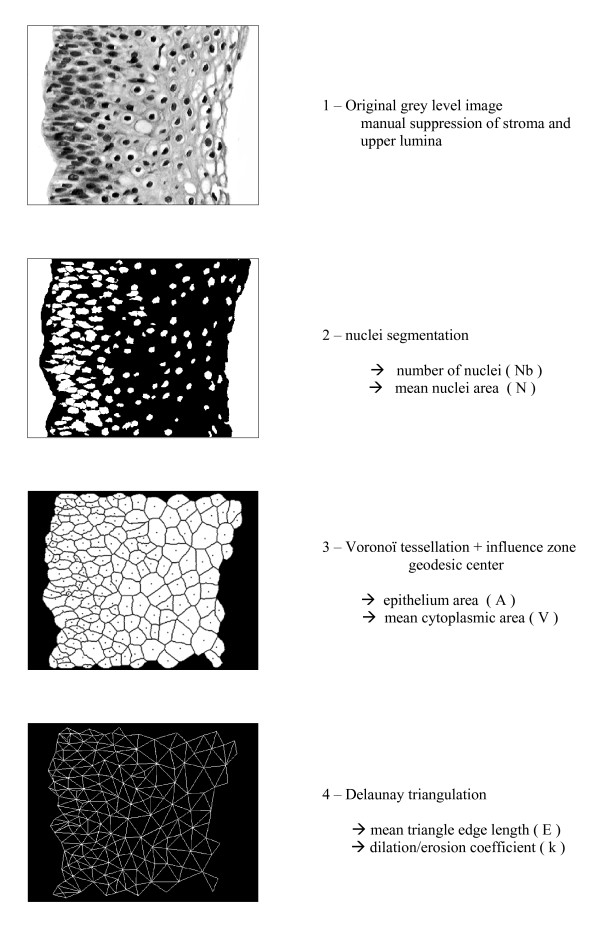
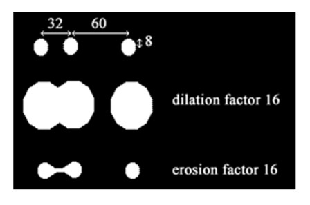
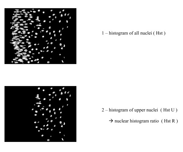
4 groups of 10 images each were selected at random from 68 consensus images coming from 80 archival cervical biopsies, normal (n = 10), CIN 1 (n = 10), CIN 2 (n = 10), CIN 3 (n = 10). Representative images of lesions were captured from the microscopic slides and were analyzed using mathematical morphology, with special reference toVoronoï tessellation and Delaunay triangulation. Epithelium surface, nuclear and cytoplasm area, triangle edge and area, total and upper nuclear index were precisely measured in each lesion, and discriminant coefficients were calculated therewith. A dilation/erosion coefficient was automatically defined using triangle edge length and nuclear radius in order to measure the epithelium ratio of differentiation. A histogram ratio was also automatically established between total nuclei and upper nuclei on top of differentiated epithelium. With the latter two ratios added to the nucleo-cytoplasmic ratio, a cervical score able to classify CIN is proposed.

RESULTS

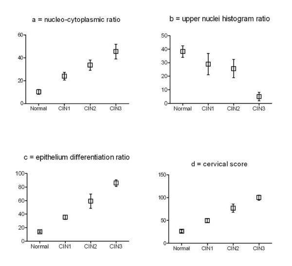
There is a quasi-linear increase of mean cervical score values between normal epithelium and CIN 3: (27) for normal epithelium, (51) for CIN 1, (78) for CIN 2 and (100) for CIN 3, with significant differences (P < 0.05).

CONCLUSION

Our results highlight the possibility of applying a cervical score for the automatic grading of CIN lesions and thereby assisting the pathologist for improvement of grading. The automatic measure of epithelium differentiation ratio appears to be a new interesting parameter in computerized image analysis of cervical lesions.

摘要

背景

尝试使用能够测量上皮分化等多个有价值参数的自动计算机系统评估宫颈内瘤变病变(CIN)的客观分级的可行性。

方法

从 80 例存档宫颈活检的 68 个共识图像中随机选择了 4 组各 10 个图像,分别为正常(n = 10)、CIN1(n = 10)、CIN2(n = 10)、CIN3(n = 10)。从显微镜载玻片上捕获病变的代表性图像,并使用数学形态学进行分析,特别参考 Voronoi 镶嵌和 Delaunay 三角剖分。在每个病变中精确测量上皮表面、核和细胞质面积、三角形边缘和面积、总核指数和上核指数,并计算相应的判别系数。通过三角形边缘长度和核半径自动定义扩张/侵蚀系数,以测量上皮分化比。还自动在上皮分化的总核和上核之间建立核质比的直方图比。将后两个比与核质比相加,提出了一种能够对 CIN 进行分类的宫颈评分。

结果

正常上皮与 CIN3 之间的平均宫颈评分值呈准线性增加:(27)为正常上皮,(51)为 CIN1,(78)为 CIN2,(100)为 CIN3,差异有统计学意义(P<0.05)。

结论

我们的结果强调了应用宫颈评分自动分级 CIN 病变的可能性,并由此协助病理学家提高分级准确性。上皮分化比的自动测量似乎是宫颈病变计算机图像分析中的一个新的有趣参数。

https://cdn.ncbi.nlm.nih.gov/pmc/blobs/d487/2819044/e7e7003eea89/1746-1596-5-7-5.jpg
https://cdn.ncbi.nlm.nih.gov/pmc/blobs/d487/2819044/b904de5cb0ae/1746-1596-5-7-1.jpg
https://cdn.ncbi.nlm.nih.gov/pmc/blobs/d487/2819044/323fd39b6a52/1746-1596-5-7-2.jpg
https://cdn.ncbi.nlm.nih.gov/pmc/blobs/d487/2819044/9dc9e10ce0e9/1746-1596-5-7-3.jpg
https://cdn.ncbi.nlm.nih.gov/pmc/blobs/d487/2819044/17f7d0eda713/1746-1596-5-7-4.jpg
https://cdn.ncbi.nlm.nih.gov/pmc/blobs/d487/2819044/e7e7003eea89/1746-1596-5-7-5.jpg

相似文献

1
Automatic measurement of epithelium differentiation and classification of cervical intraneoplasia by computerized image analysis.计算机图像分析自动测量宫颈上皮内瘤变的上皮分化和分类。
Diagn Pathol. 2010 Jan 22;5:7. doi: 10.1186/1746-1596-5-7.
2
An automated machine vision system for the histological grading of cervical intraepithelial neoplasia (CIN).一种用于宫颈上皮内瘤变(CIN)组织学分级的自动化机器视觉系统。
J Pathol. 2000 Nov;192(3):351-62. doi: 10.1002/1096-9896(2000)9999:9999<::AID-PATH708>3.0.CO;2-I.
3
False negative colposcopy is associated with thinner cervical intraepithelial neoplasia 2 and 3.阴道镜检查假阴性与较薄的宫颈上皮内瘤变2级和3级相关。
Gynecol Oncol. 2008 Jul;110(1):32-6. doi: 10.1016/j.ygyno.2008.03.003. Epub 2008 May 16.
4
Quantitative histopathological analysis of cervical intra-epithelial neoplasia sections: methodological issues.宫颈上皮内瘤变切片的定量组织病理学分析:方法学问题
Cell Oncol. 2004;26(1-2):31-43. doi: 10.1155/2004/238769.
5
Changes in the topological expression of markers of differentiation and apoptosis in defined stages of human cervical dysplasia and carcinoma.人宫颈发育异常和癌特定阶段中分化及凋亡标志物拓扑表达的变化
Gynecol Oncol. 2003 Jun;89(3):376-84. doi: 10.1016/s0090-8258(03)00061-1.
6
Correlations between nuclear/cytoplasmic area ratio and classification of cervical smears.细胞核/细胞质面积比与宫颈涂片分类之间的相关性。
J Exp Clin Cancer Res. 2001 Dec;20(4):481-6.
7
Diagnostic imaging of cervical intraepithelial neoplasia based on hematoxylin and eosin fluorescence.基于苏木精和伊红荧光的宫颈上皮内瘤变诊断成像
Diagn Pathol. 2015 Jul 25;10:119. doi: 10.1186/s13000-015-0343-8.
8
[Diagnosis and grading of cervical intraepithelial neoplasias].[宫颈上皮内瘤变的诊断与分级]
Pathologe. 2012 Mar;33(2):118-23. doi: 10.1007/s00292-011-1549-5.
9
Cytometric evidence that cervical intraepithelial neoplasia I and II are dysplasias rather than true neoplasias. An image analysis study of factors involved in the progression of cervical lesions.细胞计量学证据表明,宫颈上皮内瘤变I级和II级是发育异常而非真正的肿瘤。一项关于宫颈病变进展相关因素的图像分析研究。
Anal Quant Cytol Histol. 1992 Apr;14(2):137-47.
10
AgNOR protein quantity of cervical smears correlates with that of histological sections in cervical intraepithelial neoplasia.宫颈涂片的核仁组成区嗜银蛋白(AgNOR)含量与宫颈上皮内瘤变组织学切片的含量相关。
Eur J Histochem. 1997;41(2):105-10.

引用本文的文献

1
Deep Learning-Based Recognition of Cervical Squamous Interepithelial Lesions.基于深度学习的宫颈鳞状上皮内病变识别
Diagnostics (Basel). 2023 May 12;13(10):1720. doi: 10.3390/diagnostics13101720.
2
Gastric Pathology Image Classification Using Stepwise Fine-Tuning for Deep Neural Networks.基于深度神经网络的逐步微调进行胃病理图像分类。
J Healthc Eng. 2018 Jun 21;2018:8961781. doi: 10.1155/2018/8961781. eCollection 2018.
3
Exploring automatic prostate histopathology image Gleason grading via local structure modeling.通过局部结构建模探索前列腺组织病理学图像的自动Gleason分级

本文引用的文献

1
The reproducibility of CIN diagnoses among different pathologists: data from histology reviews from a multicenter randomized study.不同病理学家对宫颈上皮内瘤变(CIN)诊断的可重复性:来自一项多中心随机研究组织学复查的数据
Am J Clin Pathol. 2009 Jul;132(1):125-32. doi: 10.1309/AJCPBRK7D1YIUWFP.
2
Texture image analysis in differentiating malignant from benign adrenal cortical tumors in children and adults.纹理图像分析在鉴别儿童和成人肾上腺皮质肿瘤的良恶性中的应用
Anticancer Res. 2009 Aug;29(8):3365-8.
3
Theory of sampling and its application in tissue based diagnosis.
Annu Int Conf IEEE Eng Med Biol Soc. 2015;2015:2649-52. doi: 10.1109/EMBC.2015.7318936.
4
An intelligent clinical decision support system for patient-specific predictions to improve cervical intraepithelial neoplasia detection.一种用于患者特异性预测以改善宫颈上皮内瘤变检测的智能临床决策支持系统。
Biomed Res Int. 2014;2014:341483. doi: 10.1155/2014/341483. Epub 2014 Apr 9.
5
Segmentation of epidermal tissue with histopathological damage in images of haematoxylin and eosin stained human skin.苏木精和伊红染色的人类皮肤图像中具有组织病理学损伤的表皮组织分割。
BMC Med Imaging. 2014 Feb 12;14:7. doi: 10.1186/1471-2342-14-7.
6
Open source tools for management and archiving of digital microscopy data to allow integration with patient pathology and treatment information.用于管理和存档数字显微镜数据的开源工具,可与患者病理和治疗信息集成。
Diagn Pathol. 2013 Feb 12;8:22. doi: 10.1186/1746-1596-8-22.
7
Molecular characteristics and chromatin texture features in acute promyelocytic leukemia.急性早幼粒细胞白血病的分子特征和染色质纹理特征。
Diagn Pathol. 2012 Jun 28;7:75. doi: 10.1186/1746-1596-7-75.
8
Computerized texture analysis of atypical immature myeloid precursors in patients with myelodysplastic syndromes: an entity between blasts and promyelocytes.计算机纹理分析骨髓增生异常综合征患者中不典型幼稚髓系前体细胞:介于原始细胞和早幼粒细胞之间的一种实体。
Diagn Pathol. 2011 Sep 29;6:93. doi: 10.1186/1746-1596-6-93.
9
Fractal characteristics of May-Grünwald-Giemsa stained chromatin are independent prognostic factors for survival in multiple myeloma.May-Grünwald-Giemsa 染色染色质的分形特征是多发性骨髓瘤患者生存的独立预后因素。
PLoS One. 2011;6(6):e20706. doi: 10.1371/journal.pone.0020706. Epub 2011 Jun 16.
10
Fractal dimension of chromatin is an independent prognostic factor for survival in melanoma.染色质分形维数是黑色素瘤患者生存的独立预后因素。
BMC Cancer. 2010 Jun 5;10:260. doi: 10.1186/1471-2407-10-260.
采样理论及其在组织学诊断中的应用。
Diagn Pathol. 2009 Feb 16;4:6. doi: 10.1186/1746-1596-4-6.
4
Texture- and object-related automated information analysis in histological still images of various organs.不同器官组织学静态图像中与纹理和物体相关的自动信息分析
Anal Quant Cytol Histol. 2008 Dec;30(6):323-35.
5
How to measure image quality in tissue-based diagnosis (diagnostic surgical pathology).如何在组织学诊断(诊断外科病理学)中测量图像质量。
Diagn Pathol. 2008 Jul 15;3 Suppl 1(Suppl 1):S11. doi: 10.1186/1746-1596-3-S1-S11.
6
Reproducibility of the histological diagnosis of cervical dysplasia among pathologists from 4 continents.
Int J Gynecol Pathol. 2008 Jan;27(1):101-7. doi: 10.1097/pgp.0b013e31814fb1da.
7
Longitudinal evaluation of interobserver and intraobserver agreement of cervical intraepithelial neoplasia diagnosis among an experienced panel of gynecologic pathologists.在一组经验丰富的妇科病理学家中,对宫颈上皮内瘤变诊断的观察者间和观察者内一致性进行纵向评估。
Am J Surg Pathol. 2007 Dec;31(12):1854-60. doi: 10.1097/PAS.0b013e318058a544.
8
CK17 and p16 expression patterns distinguish (atypical) immature squamous metaplasia from high-grade cervical intraepithelial neoplasia (CIN III).细胞角蛋白17(CK17)和p16的表达模式可将(非典型)不成熟鳞状化生与高级别宫颈上皮内瘤变(CIN III)区分开来。
Histopathology. 2007 Apr;50(5):629-35. doi: 10.1111/j.1365-2559.2007.02652.x.
9
Kappa statistics to measure interrater and intrarater agreement for 1790 cervical biopsy specimens among twelve pathologists: qualitative histopathologic analysis and methodologic issues.用于衡量12位病理学家对1790份宫颈活检标本的评分者间和评分者内一致性的kappa统计量:定性组织病理学分析及方法学问题
Gynecol Oncol. 2005 Dec;99(3 Suppl 1):S38-52. doi: 10.1016/j.ygyno.2005.07.040. Epub 2005 Sep 23.
10
Ki67 predicts progression in early CIN: validation of a multivariate progression-risk model.Ki67可预测早期宫颈上皮内瘤变的进展:多变量进展风险模型的验证
Cell Oncol. 2004;26(1-2):13-20. doi: 10.1155/2004/108305.