Molecular Imaging Program at Stanford, Department of Radiology, School of Medicine, Stanford University, Stanford, California 94305-5105, USA.
J Nucl Med. 2010 Mar;51(3):433-40. doi: 10.2967/jnumed.109.068007. Epub 2010 Feb 11.
Targeted contrast-enhanced ultrasound imaging is increasingly being recognized as a powerful imaging tool for the detection and quantification of tumor angiogenesis at the molecular level. The purpose of this study was to develop and test a new class of targeting ligands for targeted contrast-enhanced ultrasound imaging of tumor angiogenesis with small, conformationally constrained peptides that can be coupled to the surface of ultrasound contrast agents.
Directed evolution was used to engineer a small, disulfide-constrained cystine knot (knottin) peptide that bound to alpha(v)beta(3) integrins with a low nanomolar affinity (Knottin(Integrin)). A targeted contrast-enhanced ultrasound imaging contrast agent was created by attaching Knottin(Integrin) to the shell of perfluorocarbon-filled microbubbles (MB-Knottin(Integrin)). A knottin peptide with a scrambled sequence was used to create control microbubbles (MB-Knottin(Scrambled)). The binding of MB-Knottin(Integrin) and MB-Knottin(Scrambled) to alpha(v)beta(3) integrin-positive cells and control cells was assessed in cell culture binding experiments and compared with that of microbubbles coupled to an anti-alpha(v)beta(3) integrin monoclonal antibody (MB(alphavbeta3)) and microbubbles coupled to the peptidomimetic agent c(RGDfK) (MB(cRGD)). The in vivo imaging signals of contrast-enhanced ultrasound with the different types of microbubbles were quantified in 42 mice bearing human ovarian adenocarcinoma xenograft tumors by use of a high-resolution 40-MHz ultrasound system.
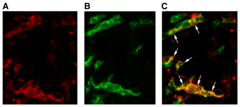
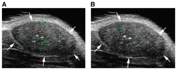
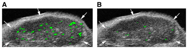
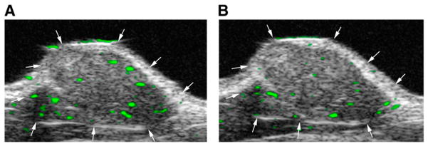
MB-Knottin(Integrin) attached significantly more to alpha(v)beta(3) integrin-positive cells (1.76 +/- 0.49 [mean +/- SD] microbubbles per cell) than to control cells (0.07 +/- 0.006). Control MB-Knottin(Scrambled) adhered less to alpha(v)beta(3) integrin-positive cells (0.15 +/- 0.12) than MB-Knottin(Integrin). After blocking of integrins, the attachment of MB-Knottin(Integrin) to alpha(v)beta(3) integrin-positive cells decreased significantly. The in vivo ultrasound imaging signal was significantly higher after the administration of MB-Knottin(Integrin) than after the administration of MB(alphavbeta3) or control MB-Knottin(Scrambled). After in vivo blocking of integrin receptors, the imaging signal after the administration of MB-Knottin(Integrin) decreased significantly (by 64%). The imaging signals after the administration of MB-Knottin(Integrin) were not significantly different in the groups of tumor-bearing mice imaged with MB-Knottin(Integrin) and with MB(cRGD). Ex vivo immunofluorescence confirmed integrin expression on endothelial cells of human ovarian adenocarcinoma xenograft tumors.
Integrin-binding knottin peptides can be conjugated to the surface of microbubbles and used for in vivo targeted contrast-enhanced ultrasound imaging of tumor angiogenesis. Our results demonstrate that microbubbles conjugated to small peptide-targeting ligands provide imaging signals higher than those provided by a large antibody molecule.
本研究旨在开发并测试一类新的靶向配体,用于靶向对比增强超声成像检测肿瘤血管生成,使用的靶向配体是可以偶联到超声造影剂表面的小型、构象受限的肽。
通过定向进化工程,构建了一种与αvβ3 整合素具有低纳摩尔亲和力的小型二硫键约束半胱氨酸结(knottin)肽(Knottin(Integrin))。通过将 Knottin(Integrin)偶联到全氟碳填充微泡(MB-Knottin(Integrin))的壳上,创建了一种靶向对比增强超声成像造影剂。用具有乱序序列的 knottin 肽创建了对照微泡(MB-Knottin(Scrambled))。在细胞培养结合实验中评估了 MB-Knottin(Integrin)和 MB-Knottin(Scrambled)与 αvβ3 整合素阳性细胞和对照细胞的结合情况,并与与抗 αvβ3 整合素单克隆抗体偶联的微泡(MB(alphavbeta3))和与肽模拟物 c(RGDfK)偶联的微泡(MB(cRGD))进行了比较。通过使用高分辨率 40MHz 超声系统,在 42 只荷有人卵巢腺癌异种移植肿瘤的小鼠中定量评估了不同类型微泡的对比增强超声成像信号。
MB-Knottin(Integrin)与 αvβ3 整合素阳性细胞的结合明显多于与对照细胞的结合(每个细胞 1.76 ± 0.49 [平均值 ± SD]个微泡)。对照 MB-Knottin(Scrambled)与 αvβ3 整合素阳性细胞的结合(0.15 ± 0.12)少于 MB-Knottin(Integrin)。整合素被阻断后,MB-Knottin(Integrin)与 αvβ3 整合素阳性细胞的结合明显减少。MB-Knottin(Integrin)给药后的超声成像信号明显高于 MB(alphavbeta3)或对照 MB-Knottin(Scrambled)给药后的信号。体内阻断整合素受体后,MB-Knottin(Integrin)给药后的成像信号明显降低(64%)。用 MB-Knottin(Integrin)和 MB(cRGD)成像的荷瘤小鼠组之间的成像信号无明显差异。体外免疫荧光证实了人卵巢腺癌异种移植肿瘤内皮细胞的整合素表达。
整合素结合 knottin 肽可偶联到微泡表面,用于体内靶向对比增强超声成像检测肿瘤血管生成。我们的结果表明,与大抗体分子相比,偶联小肽靶向配体的微泡提供了更高的成像信号。