• 文献检索
  • 文档翻译
  • 深度研究
  • 学术资讯
  • Suppr Zotero 插件Zotero 插件
  • 邀请有礼
  • 套餐&价格
  • 历史记录
应用&插件
Suppr Zotero 插件Zotero 插件浏览器插件Mac 客户端Windows 客户端微信小程序
定价
高级版会员购买积分包购买API积分包
服务
文献检索文档翻译深度研究API 文档MCP 服务
关于我们
关于 Suppr公司介绍联系我们用户协议隐私条款
关注我们

Suppr 超能文献

核心技术专利:CN118964589B侵权必究
粤ICP备2023148730 号-1Suppr @ 2026

文献检索

告别复杂PubMed语法,用中文像聊天一样搜索,搜遍4000万医学文献。AI智能推荐,让科研检索更轻松。

立即免费搜索

文件翻译

保留排版,准确专业,支持PDF/Word/PPT等文件格式,支持 12+语言互译。

免费翻译文档

深度研究

AI帮你快速写综述,25分钟生成高质量综述,智能提取关键信息,辅助科研写作。

立即免费体验

在兔实验性动脉分叉处观察到的光镜和扫描电镜变化。

Light and scanning electron microscopic changes observed in experimental arterial forks of rabbits.

作者信息

Stehbens W E, Martin B J, Delahunt B

机构信息

Department of Pathology, Wellington School of Medicine, New Zealand.

出版信息

Int J Exp Pathol. 1991 Apr;72(2):183-93.

PMID:2015201
原文链接:https://pmc.ncbi.nlm.nih.gov/articles/PMC2002305/
Abstract

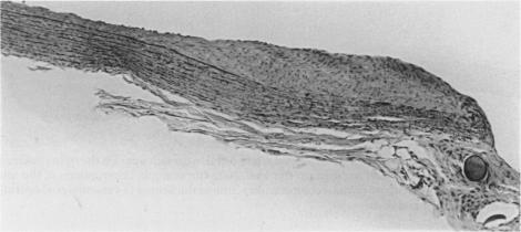
Experimental arterial forks were fashioned by anastomosing one common carotid artery to the contralateral vessel by microvascular surgery in rabbits. In one rabbit group the forks were examined histologically by the serial section technique from 5.5 to 25 months postoperatively. The second group was used for scanning electron microscopy of the arterial endothelial surface from 1 to 257 days post-operatively. Intimal proliferation was observed at the lateral angles histologically at sites comparable to those where intimal proliferation occurs spontaneously in human infants. In addition, disruption of the internal elastic lamina with minimal intimal proliferation occurred at the sides of the neo-apex, mostly in the transposed artery. These disruptions corresponded to predominantly transversely orientated tears of the internal elastic lamina from 8 days post-operatively in the scanning electron microscopic study. They were similar to the early atrophic changes preceding berry aneurysm formation in human cerebral arterial forks. The results indicate that both intimal pads (cushions) and elastic tears can be haemodynamically induced.

摘要

通过微血管手术将一只家兔的颈总动脉与对侧血管吻合,制作实验性动脉分叉模型。在一组家兔中,术后5.5至25个月采用连续切片技术对分叉处进行组织学检查。第二组用于术后1至257天对动脉内皮表面进行扫描电子显微镜检查。组织学观察发现,在与人类婴儿自发发生内膜增生部位相当的外侧角处出现了内膜增生。此外,在新顶端两侧出现了内弹性膜破坏且内膜增生轻微的情况,主要发生在移位动脉中。在扫描电子显微镜研究中,这些破坏对应于术后8天起内弹性膜主要呈横向的撕裂。它们类似于人类脑动脉分叉处浆果样动脉瘤形成之前的早期萎缩性变化。结果表明,内膜垫(衬垫)和弹性撕裂均可由血流动力学诱导产生。

https://cdn.ncbi.nlm.nih.gov/pmc/blobs/7941/2002305/ed588393e35c/ijexpath00026-0082-b.jpg
https://cdn.ncbi.nlm.nih.gov/pmc/blobs/7941/2002305/8b65755c7e9c/ijexpath00026-0078-a.jpg
https://cdn.ncbi.nlm.nih.gov/pmc/blobs/7941/2002305/ef998799e019/ijexpath00026-0078-b.jpg
https://cdn.ncbi.nlm.nih.gov/pmc/blobs/7941/2002305/03258d109bae/ijexpath00026-0079-a.jpg
https://cdn.ncbi.nlm.nih.gov/pmc/blobs/7941/2002305/c197ce01ab68/ijexpath00026-0080-a.jpg
https://cdn.ncbi.nlm.nih.gov/pmc/blobs/7941/2002305/b6fa4aa5a93a/ijexpath00026-0080-b.jpg
https://cdn.ncbi.nlm.nih.gov/pmc/blobs/7941/2002305/bee034890977/ijexpath00026-0081-a.jpg
https://cdn.ncbi.nlm.nih.gov/pmc/blobs/7941/2002305/7e9ca144f7e6/ijexpath00026-0082-a.jpg
https://cdn.ncbi.nlm.nih.gov/pmc/blobs/7941/2002305/ed588393e35c/ijexpath00026-0082-b.jpg

相似文献

1
Light and scanning electron microscopic changes observed in experimental arterial forks of rabbits.在兔实验性动脉分叉处观察到的光镜和扫描电镜变化。
Int J Exp Pathol. 1991 Apr;72(2):183-93.
2
Endothelium and elastic tears in the afferent arteries of experimental arteriovenous fistulae in rabbits.兔实验性动静脉瘘传入动脉的内皮及弹性组织撕裂
Int J Exp Pathol. 1992 Aug;73(4):405-16.
3
Haemodynamically-induced intimal tears in experimental U-shaped arterial loops as seen by scanning electron microscopy.扫描电子显微镜观察到的实验性U形动脉袢中血流动力学诱导的内膜撕裂。
Br J Exp Pathol. 1985 Oct;66(5):577-84.
4
[Study of the elastic skeleton of intracranial arteries in animal and human vessels and experimentally induced cerebral aneurysms].[动物和人体血管以及实验性诱发脑动脉瘤中颅内动脉弹性骨架的研究]
Nihon Geka Hokan. 1991 Jan 1;60(1):13-24.
5
Ultrastructural changes in arteries proximal to short-term experimental carotid-jugular arteriovenous fistulae in rabbits.兔短期实验性颈总动脉 - 颈外静脉动静脉瘘近端动脉的超微结构变化
Int J Exp Pathol. 1994 Jun;75(3):225-32.
6
Scanning electron microscopic study of hemodynamically induced tears in the internal elastic lamina of rabbit arteries.兔动脉内弹性膜血流动力学诱导撕裂的扫描电子显微镜研究。
Pathology. 1989 Jul;21(3):207-12. doi: 10.3109/00313028909061060.
7
Scanning electron-microscopic study of experimentally induced intimal tears in rabbit arteries.兔动脉实验性内膜撕裂的扫描电子显微镜研究。
Atherosclerosis. 1983 Nov;49(2):119-26. doi: 10.1016/0021-9150(83)90189-2.
8
Early changes of experimentally induced cerebral aneurysms in rats. Light-microscopic study.大鼠实验性诱导脑动脉瘤的早期变化。光学显微镜研究。
Am J Pathol. 1986 Sep;124(3):399-404.
9
A scanning electron microscopy study of the endothelial folds in normal and anastomosed common carotid arteries in albino rats.白化大鼠正常及吻合后的颈总动脉内皮皱襞的扫描电子显微镜研究
Br J Exp Pathol. 1975 Aug;56(4):329-37.
10
Vascular clips have no significant effect on the cellular proliferation, intimal changes, or peak systolic velocity at anastomoses in rabbit vein grafts.血管夹对兔静脉移植物吻合处的细胞增殖、内膜变化或收缩期峰值流速无显著影响。
J Surg Res. 2000 Jul;92(1):29-35. doi: 10.1006/jsre.2000.5843.

引用本文的文献

1
Physical factors effecting cerebral aneurysm pathophysiology.影响颅内动脉瘤病理生理学的物理因素。
Ann Biomed Eng. 2013 Jul;41(7):1347-65. doi: 10.1007/s10439-013-0800-z. Epub 2013 Apr 3.
2
Ultrastructural changes in arteries proximal to short-term experimental carotid-jugular arteriovenous fistulae in rabbits.兔短期实验性颈总动脉 - 颈外静脉动静脉瘘近端动脉的超微结构变化
Int J Exp Pathol. 1994 Jun;75(3):225-32.
3
Endothelium and elastic tears in the afferent arteries of experimental arteriovenous fistulae in rabbits.兔实验性动静脉瘘传入动脉的内皮及弹性组织撕裂

本文引用的文献

1
Histopathology of cerebral aneurysms.脑动脉瘤的组织病理学
Arch Neurol. 1963 Mar;8:272-85. doi: 10.1001/archneur.1963.00460030056005.
2
Focal intimal proliferation in the cerebral arteries.脑动脉局灶性内膜增生。
Am J Pathol. 1960 Mar;36(3):289-301.
3
A study of the influence of steroid hormones, hemodynamics, and environmental factors on the formation of small "spontaneous" lesions in the rat caudal artery.
Lab Invest. 1980 Nov;43(5):471-9.
Int J Exp Pathol. 1992 Aug;73(4):405-16.
4
Changes in the cross-sectional area of the arterial fork.动脉分叉处横截面积的变化。
Angiology. 1974 Oct;25(9):561-75. doi: 10.1177/000331977402500902.
5
Calcifications of iliac arteries in newborns and infants.新生儿和婴儿髂动脉钙化
Arch Dis Child. 1972 Jun;47(253):364-72. doi: 10.1136/adc.47.253.364.
6
Haemodynamically-induced intimal tears in experimental U-shaped arterial loops as seen by scanning electron microscopy.扫描电子显微镜观察到的实验性U形动脉袢中血流动力学诱导的内膜撕裂。
Br J Exp Pathol. 1985 Oct;66(5):577-84.
7
An animal model of cerebral aneurysms.脑动脉瘤的动物模型。
Neuropathol Appl Neurobiol. 1987 Mar-Apr;13(2):77-90. doi: 10.1111/j.1365-2990.1987.tb00173.x.
8
Scanning electron microscopic investigation of the afferent arteries of experimental femoral arteriovenous fistulae in rabbits.兔实验性股动静脉瘘传入动脉的扫描电子显微镜研究。
Pathology. 1987 Jan;19(1):22-7. doi: 10.3109/00313028709065130.
9
Early membrane damage during ischemia in rat heart.大鼠心脏缺血期间的早期膜损伤。
Exp Mol Pathol. 1986 Feb;44(1):1-6. doi: 10.1016/0014-4800(86)90027-4.