• 文献检索
  • 文档翻译
  • 深度研究
  • 学术资讯
  • Suppr Zotero 插件Zotero 插件
  • 邀请有礼
  • 套餐&价格
  • 历史记录
应用&插件
Suppr Zotero 插件Zotero 插件浏览器插件Mac 客户端Windows 客户端微信小程序
定价
高级版会员购买积分包购买API积分包
服务
文献检索文档翻译深度研究API 文档MCP 服务
关于我们
关于 Suppr公司介绍联系我们用户协议隐私条款
关注我们

Suppr 超能文献

核心技术专利:CN118964589B侵权必究
粤ICP备2023148730 号-1Suppr @ 2026

文献检索

告别复杂PubMed语法,用中文像聊天一样搜索,搜遍4000万医学文献。AI智能推荐,让科研检索更轻松。

立即免费搜索

文件翻译

保留排版,准确专业,支持PDF/Word/PPT等文件格式,支持 12+语言互译。

免费翻译文档

深度研究

AI帮你快速写综述,25分钟生成高质量综述,智能提取关键信息,辅助科研写作。

立即免费体验

扫描电子显微镜观察到的实验性U形动脉袢中血流动力学诱导的内膜撕裂。

Haemodynamically-induced intimal tears in experimental U-shaped arterial loops as seen by scanning electron microscopy.

作者信息

Greenhill N S, Stehbens W E

出版信息

Br J Exp Pathol. 1985 Oct;66(5):577-84.

PMID:4063161
原文链接:https://pmc.ncbi.nlm.nih.gov/articles/PMC2042049/
Abstract

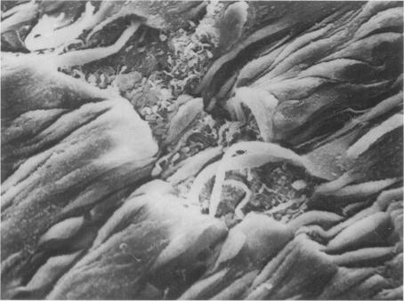
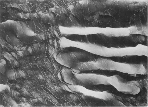
Experimentally-induced U-shaped carotid loops, simulating arterial tortuosities and kinks were examined by scanning electron microscopy to seek flow-induced changes in the intimal surface. In 14 New Zealand white rabbits carotid arterial transplants were fashioned into U-shaped loops by microvascular surgery. The rabbits were sacrificed from 4 to 226 days post-operatively. Tears in the internal elastic lamina occurred in all loops from 5 days post-operatively and were predominantly transverse and localised about the greater curvature of the bends of each loop. Though initially denuded, all tears appeared endothelialised after 6 days, coalescing later as they increased in size and extent. In older animals only islands of wrinkled internal elastic lamina remained at those sites. Endothelial cells in the tears were small, numerous and polyhedral with raised nuclei. The lesser curvature of the three bends in the loops displayed some irregular wrinkling of the internal elastic lamina. In the three oldest animals a few longitudinal tears were observed on the lesser curvature of the main bend. The specific localization of intimal tears supports the concept that they were hemodynamically induced.

摘要

通过扫描电子显微镜检查实验诱导的U形颈动脉袢,模拟动脉迂曲和扭结,以寻找血流诱导的内膜表面变化。在14只新西兰白兔中,通过微血管手术将颈动脉移植制成U形袢。术后4至226天处死兔子。术后5天所有袢的内弹性膜均出现撕裂,主要为横向撕裂,且局限于每个袢弯曲的大曲率处。虽然最初内膜剥脱,但所有撕裂在6天后似乎都有内皮化,随着其大小和范围的增加,后来融合在一起。在老年动物中,只有起皱的内弹性膜岛留在这些部位。撕裂处的内皮细胞小、数量多且呈多面体,细胞核凸起。袢中三个弯曲的小曲率处显示内弹性膜有一些不规则的起皱。在三只年龄最大的动物中,在主弯曲的小曲率处观察到一些纵向撕裂。内膜撕裂的特定定位支持了它们是由血流动力学诱导的这一概念。

https://cdn.ncbi.nlm.nih.gov/pmc/blobs/a969/2042049/d59f4b173d19/brjexppathol00089-0072-a.jpg
https://cdn.ncbi.nlm.nih.gov/pmc/blobs/a969/2042049/ea8dbb90b56b/brjexppathol00089-0069-a.jpg
https://cdn.ncbi.nlm.nih.gov/pmc/blobs/a969/2042049/3cee832b2f47/brjexppathol00089-0070-a.jpg
https://cdn.ncbi.nlm.nih.gov/pmc/blobs/a969/2042049/9786ccda08b4/brjexppathol00089-0070-b.jpg
https://cdn.ncbi.nlm.nih.gov/pmc/blobs/a969/2042049/fc51fe37bb3c/brjexppathol00089-0071-a.jpg
https://cdn.ncbi.nlm.nih.gov/pmc/blobs/a969/2042049/d59f4b173d19/brjexppathol00089-0072-a.jpg

相似文献

1
Haemodynamically-induced intimal tears in experimental U-shaped arterial loops as seen by scanning electron microscopy.扫描电子显微镜观察到的实验性U形动脉袢中血流动力学诱导的内膜撕裂。
Br J Exp Pathol. 1985 Oct;66(5):577-84.
2
Light and scanning electron microscopic changes observed in experimental arterial forks of rabbits.在兔实验性动脉分叉处观察到的光镜和扫描电镜变化。
Int J Exp Pathol. 1991 Apr;72(2):183-93.
3
Endothelium and elastic tears in the afferent arteries of experimental arteriovenous fistulae in rabbits.兔实验性动静脉瘘传入动脉的内皮及弹性组织撕裂
Int J Exp Pathol. 1992 Aug;73(4):405-16.
4
Scanning electron-microscopic study of experimentally induced intimal tears in rabbit arteries.兔动脉实验性内膜撕裂的扫描电子显微镜研究。
Atherosclerosis. 1983 Nov;49(2):119-26. doi: 10.1016/0021-9150(83)90189-2.
5
Scanning electron microscopic study of hemodynamically induced tears in the internal elastic lamina of rabbit arteries.兔动脉内弹性膜血流动力学诱导撕裂的扫描电子显微镜研究。
Pathology. 1989 Jul;21(3):207-12. doi: 10.3109/00313028909061060.
6
Experimental arterial loops and arterial atrophy.实验性动脉袢与动脉萎缩
Exp Mol Pathol. 1986 Apr;44(2):177-89. doi: 10.1016/0014-4800(86)90068-7.
7
Ultrastructural changes in arteries proximal to short-term experimental carotid-jugular arteriovenous fistulae in rabbits.兔短期实验性颈总动脉 - 颈外静脉动静脉瘘近端动脉的超微结构变化
Int J Exp Pathol. 1994 Jun;75(3):225-32.
8
Scanning electron microscopic investigation of the afferent arteries of experimental femoral arteriovenous fistulae in rabbits.兔实验性股动静脉瘘传入动脉的扫描电子显微镜研究。
Pathology. 1987 Jan;19(1):22-7. doi: 10.3109/00313028709065130.
9
Superficial intimal injury of the rabbit carotid artery induced by distilled water.蒸馏水诱导兔颈动脉内膜浅层损伤。
Virchows Arch B Cell Pathol Incl Mol Pathol. 1986;51(3):197-205. doi: 10.1007/BF02899029.
10
Scanning electron-microscopic study of the inner surface of experimental aneurysms in rabbits.兔实验性动脉瘤内表面的扫描电子显微镜研究。
Atherosclerosis. 1982 Dec;45(3):319-30. doi: 10.1016/0021-9150(82)90233-7.

引用本文的文献

1
Extracranial carotid arteriopathy in stroke-free children with sickle cell anemia: detection by submandibular Doppler sonography.镰状细胞贫血无卒中患儿的颅外颈动脉病变:通过下颌下多普勒超声检测
Pediatr Radiol. 2014 May;44(5):587-96. doi: 10.1007/s00247-014-2880-9. Epub 2014 Mar 6.
2
A steering model of endothelial sheet migration recapitulates monolayer integrity and directed collective migration.内皮细胞片层迁移的导向模型再现了单层完整性和定向集体迁移。
Mol Cell Biol. 2011 Jan;31(2):342-50. doi: 10.1128/MCB.00800-10. Epub 2010 Oct 25.
3
Regression of a Flow-Related Ophthalmic Artery Aneurysm After Treatment of a Frontal DAVS. A Case Report.

本文引用的文献

1
A hemodynamic concept of atherosclerosis, with particular reference to coronary occlusion.动脉粥样硬化的血流动力学概念,尤其涉及冠状动脉闭塞。
AMA Arch Intern Med. 1957 Mar;99(3):418-27. doi: 10.1001/archinte.1957.00260030100010.
2
Scanning electron microscopy of endothelium around an experimental stenosis in the rabbit aorta using a new casting material.使用一种新型铸型材料对兔主动脉实验性狭窄周围内皮进行扫描电子显微镜检查。
Atherosclerosis. 1982 Apr;42(2-3):299-318. doi: 10.1016/0021-9150(82)90158-7.
3
[Calcinosis and the system of gaps in the internal elastic membrane of tortuous splenic arteries].
额叶硬脑膜动静脉瘘治疗后血流相关性眼动脉动脉瘤的消退。病例报告。
Interv Neuroradiol. 2004 Sep 30;10(3):265-8. doi: 10.1177/159101990401000310. Epub 2005 Jan 5.
4
Ultrastructural changes in arteries proximal to short-term experimental carotid-jugular arteriovenous fistulae in rabbits.兔短期实验性颈总动脉 - 颈外静脉动静脉瘘近端动脉的超微结构变化
Int J Exp Pathol. 1994 Jun;75(3):225-32.
5
Light and scanning electron microscopic changes observed in experimental arterial forks of rabbits.在兔实验性动脉分叉处观察到的光镜和扫描电镜变化。
Int J Exp Pathol. 1991 Apr;72(2):183-93.
6
The rat testicular artery: a model of spontaneous aneurysmal-like structure formation.大鼠睾丸动脉:一种自发性动脉瘤样结构形成的模型。
Am J Pathol. 1992 Nov;141(5):1053-61.
7
Endothelium and elastic tears in the afferent arteries of experimental arteriovenous fistulae in rabbits.兔实验性动静脉瘘传入动脉的内皮及弹性组织撕裂
Int J Exp Pathol. 1992 Aug;73(4):405-16.
[弯曲脾动脉内弹性膜中的钙质沉着与间隙系统]
Virchows Arch A Pathol Pathol Anat. 1968;345(3):292-8.
4
Calcifications of iliac arteries in newborns and infants.新生儿和婴儿髂动脉钙化
Arch Dis Child. 1972 Jun;47(253):364-72. doi: 10.1136/adc.47.253.364.
5
Preparation of vascular endothelium for scanning electron microscopy: a comparison of the effects of perfusion and immersion fixation.用于扫描电子显微镜检查的血管内皮制备:灌注固定和浸入固定效果的比较
J Microsc. 1975 Dec;105(3):309-13. doi: 10.1111/j.1365-2818.1975.tb04065.x.
6
The role of hemodynamic factors in arterial wall thickening in the rat.血流动力学因素在大鼠动脉壁增厚中的作用。
Atherosclerosis. 1978 Mar;29(3):363-71. doi: 10.1016/0021-9150(78)90083-7.