Bioinformatics Institute (A-STAR), Matrix, Singapore.
BMC Genomics. 2010 Feb 10;11 Suppl 1(Suppl 1):S5. doi: 10.1186/1471-2164-11-S1-S5.
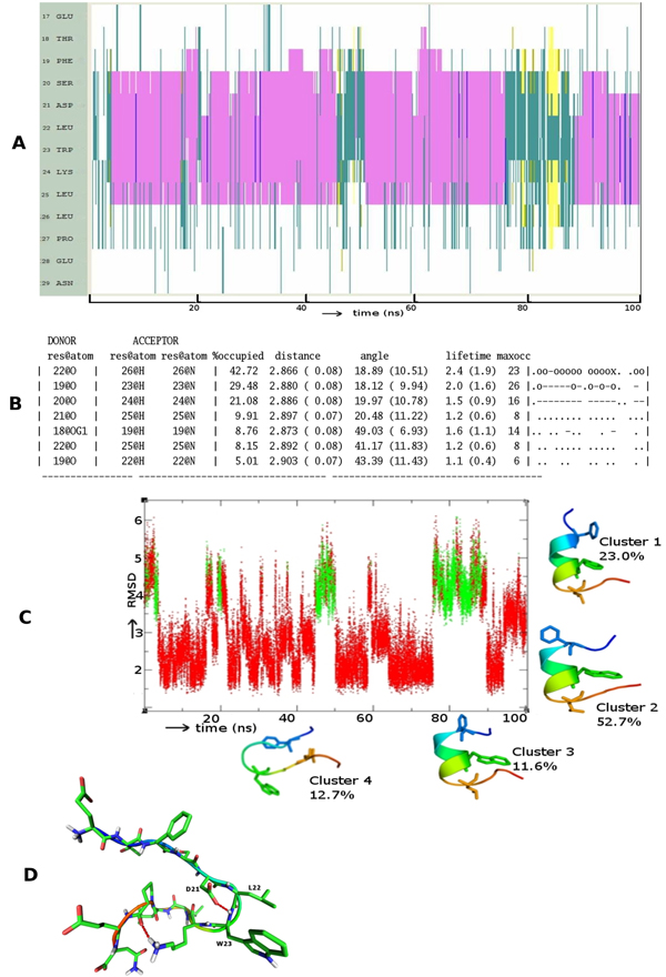
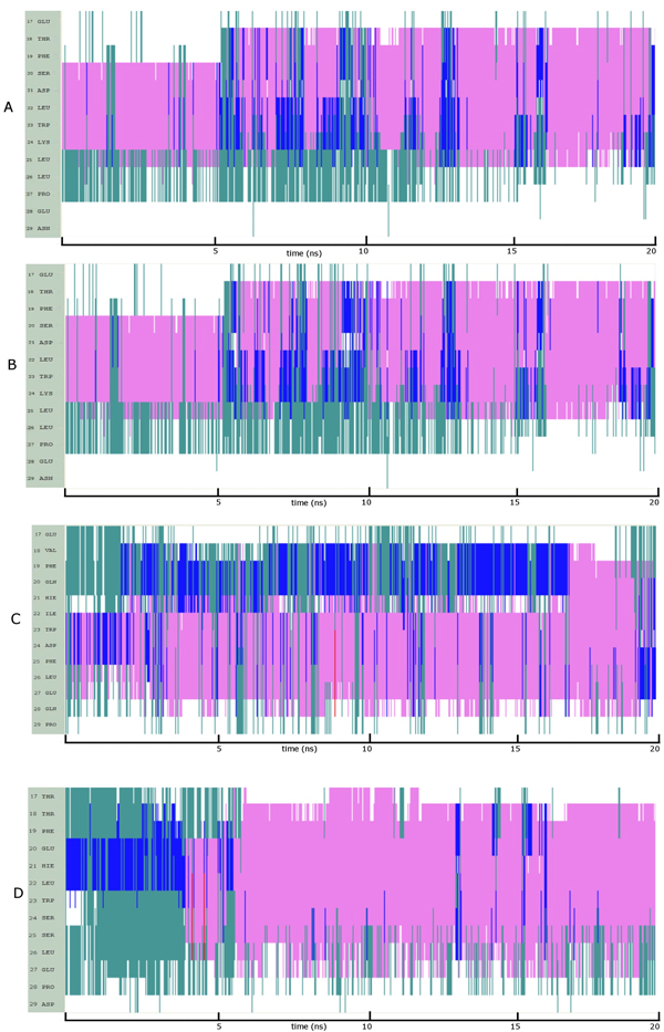
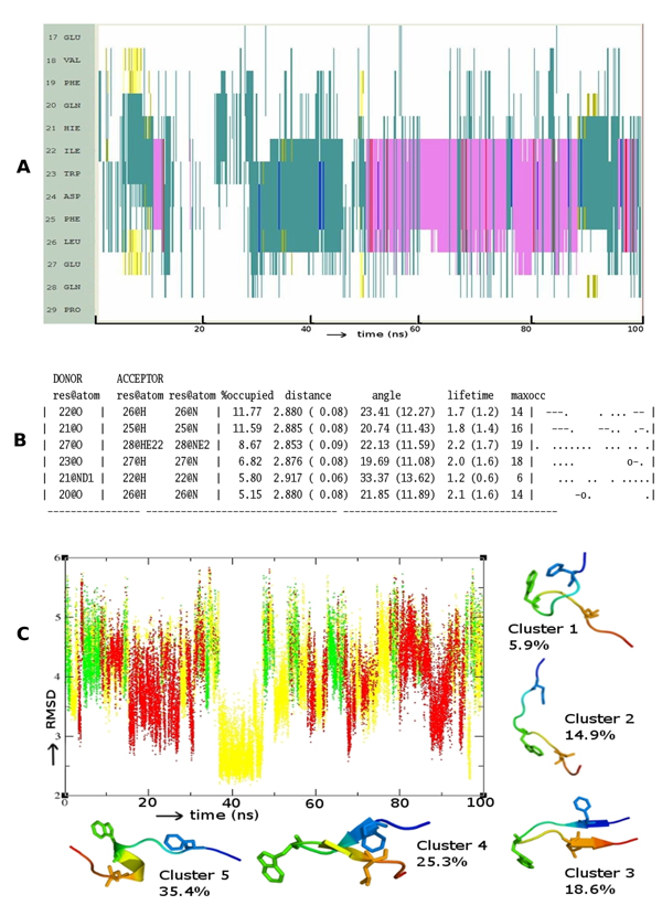
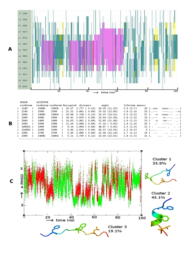
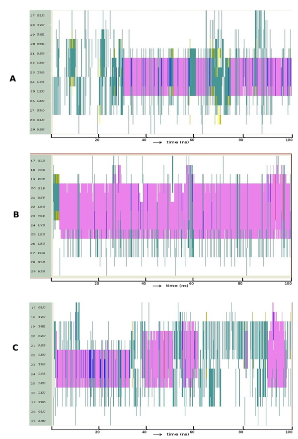
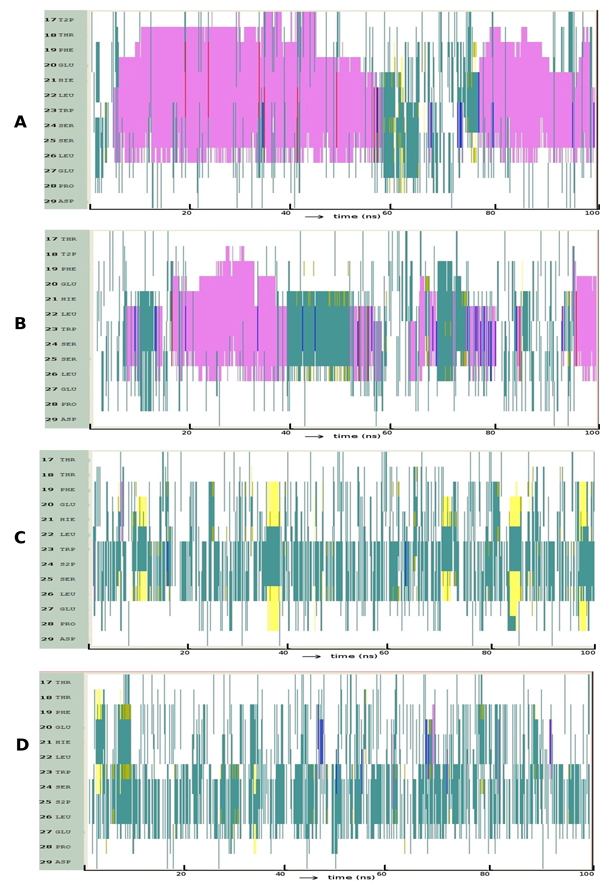
The N terminal transactivation domain of p53 is regulated by ligases and coactivator proteins. The functional conformation of this region appears to be an alpha helix which is necessary for its appropriate interactions with several proteins including MDM2 and p300. Folding simulation studies have been carried out to examine the propensity and stability of this region and are used to understand the differences between the family members with the ease of helix formation following the order p53 > p73 > p63. It is clear that hydrophobic clusters control the kinetics of helix formation, while electrostatic interactions control the thermodynamic stability of the helix. Differences in these interactions between the family members may partially account for the differential binding to, and regulation by, MDM2 (and MDMX). Phosphorylations of the peptides further modulate the stability of the helix and control associations with partner proteins.
p53 的 N 端反式激活结构域受连接酶和辅激活蛋白的调控。该区域的功能构象似乎是一个α螺旋,这对于其与包括 MDM2 和 p300 在内的几种蛋白质的适当相互作用是必需的。已进行折叠模拟研究以检查该区域的倾向性和稳定性,并用于理解家族成员之间的差异,其易于形成螺旋的顺序为 p53 > p73 > p63。显然,疏水区簇控制着螺旋形成的动力学,而静电相互作用则控制着螺旋的热力学稳定性。家族成员之间这些相互作用的差异可能部分解释了它们与 MDM2(和 MDMX)的不同结合和调节方式。肽的磷酸化进一步调节螺旋的稳定性并控制与伴侣蛋白的关联。