Digirad Corporation, 13950 Stowe Drive, Poway, CA 92064, USA.
J Nucl Cardiol. 2010 Jun;17(3):459-69. doi: 10.1007/s12350-010-9204-8. Epub 2010 Feb 19.
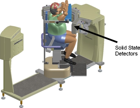
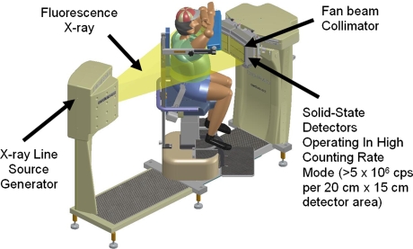
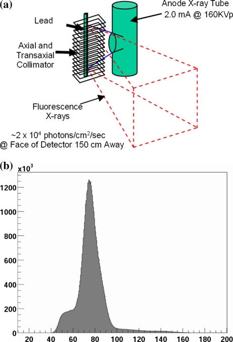
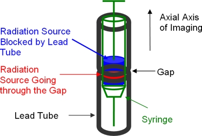
We developed a cardiac SPECT system (X-ACT) with low dose volume CT transmission-based attenuation correction (AC). Three solid-state detectors are configured to form a triple-head system for emission scans and reconfigured to form a 69-cm field-of-view detector arc for transmission scans. A near mono-energetic transmission line source is produced from the collimated fluorescence x-ray emitted from a lead target when the target is illuminated by a narrow polychromatic x-ray beam from an x-ray tube. Transmission scans can be completed in 1 min with insignificant patient dose (deep dose equivalent <5 muSv).
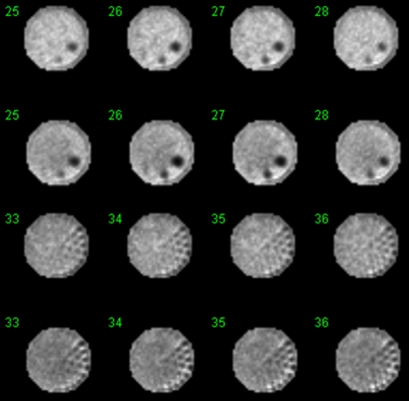
We used phantom studies to evaluate (1) the accuracy of the reconstructed attenuation maps, (2) the effect of AC on image uniformity, and (3) the effect of AC on defect contrast (DC). The phantoms we used included an ACR phantom, an anthropomorphic phantom with a uniform cardiac insert, and an anthropomorphic phantom with two defects in the cardiac insert.
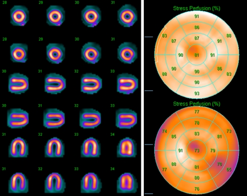
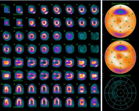
The reconstructed attenuation coefficient of water at 140 keV was .150 +/- .003/cm in the uniform region of the ACR phantom, .151 +/- .003/cm and .151 +/- .002/cm in the liver and cardiac regions of the anthropomorphic phantom. The ACR phantom images with AC showed correction of the bowing effect due to attenuation in the images without AC (NC). The 17-segment scores of the images of the uniform cardiac insert were 78.3 +/- 6.5 before and 87.9 +/- 3.3 after AC (average +/- standard deviation). The inferior-to-anterior wall ratio and the septal-to-lateral wall ratio were .99 and 1.16 before and 1.02 and 1.00 after AC. The DC of the two defects was .528 and .156 before and .628 and .173 after AC.
The X-ACT system generated accurate attenuation maps with 1-minute transmission scans. AC improved image quality and uniformity over NC.
我们开发了一种具有低剂量体积 CT 透射式衰减校正(AC)的心脏 SPECT 系统(X-ACT)。三个固态探测器配置为形成用于发射扫描的三头系统,并重新配置为形成用于透射扫描的 69cm 视场探测器弧形。当目标被来自 X 射线管的窄多色 X 射线束照亮时,从铅目标发出的准单能荧光 X 射线产生近单能透射线源。透射扫描可以在 1 分钟内完成,患者剂量微不足道(深部剂量当量<5μSv)。
我们使用体模研究来评估(1)重建衰减图的准确性,(2)AC 对图像均匀性的影响,以及(3)AC 对缺陷对比度(DC)的影响。我们使用的体模包括 ACR 体模、带有均匀心脏插件的拟人化体模和带有两个心脏插件缺陷的拟人化体模。
在 ACR 体模的均匀区域,水的重建衰减系数在 140keV 下为 0.150±0.003/cm,在肝脏和心脏区域的拟人化体模中为 0.151±0.003/cm 和 0.151±0.002/cm。AC 后的图像校正了 NC 中由于衰减引起的弯曲效应。均匀心脏插件图像的 17 节评分在 AC 前为 78.3±6.5,AC 后为 87.9±3.3(平均值±标准差)。下壁到前壁比和间隔到侧壁比分别为 AC 前的 0.99 和 1.16,AC 后的 1.02 和 1.00。两个缺陷的 DC 在 AC 前分别为 0.528 和 0.156,AC 后分别为 0.628 和 0.173。
X-ACT 系统在 1 分钟的透射扫描中生成了准确的衰减图。AC 提高了 NC 后的图像质量和均匀性。