• 文献检索
  • 文档翻译
  • 深度研究
  • 学术资讯
  • Suppr Zotero 插件Zotero 插件
  • 邀请有礼
  • 套餐&价格
  • 历史记录
应用&插件
Suppr Zotero 插件Zotero 插件浏览器插件Mac 客户端Windows 客户端微信小程序
定价
高级版会员购买积分包购买API积分包
服务
文献检索文档翻译深度研究API 文档MCP 服务
关于我们
关于 Suppr公司介绍联系我们用户协议隐私条款
关注我们

Suppr 超能文献

核心技术专利:CN118964589B侵权必究
粤ICP备2023148730 号-1Suppr @ 2026

文献检索

告别复杂PubMed语法,用中文像聊天一样搜索,搜遍4000万医学文献。AI智能推荐,让科研检索更轻松。

立即免费搜索

文件翻译

保留排版,准确专业,支持PDF/Word/PPT等文件格式,支持 12+语言互译。

免费翻译文档

深度研究

AI帮你快速写综述,25分钟生成高质量综述,智能提取关键信息,辅助科研写作。

立即免费体验

大鼠脑内皮细胞是锰毒性的靶细胞。

Rat brain endothelial cells are a target of manganese toxicity.

机构信息

i-Med-UL, Faculdade de Farmácia, Universidade de Lisboa, Lisbon, Portugal.

出版信息

Brain Res. 2010 Apr 22;1326:152-61. doi: 10.1016/j.brainres.2010.02.016. Epub 2010 Feb 17.

DOI:10.1016/j.brainres.2010.02.016
PMID:20170646
原文链接:https://pmc.ncbi.nlm.nih.gov/articles/PMC2849855/
Abstract

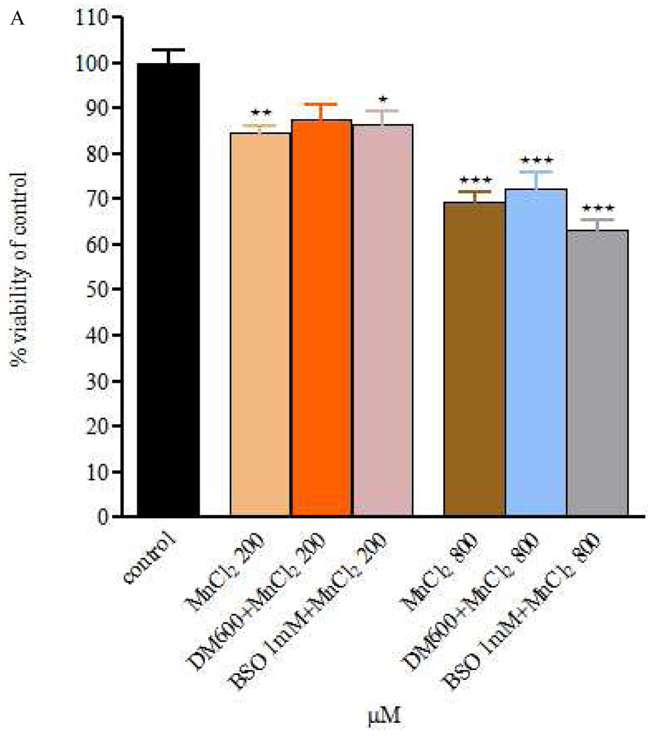
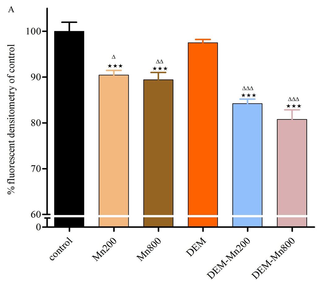
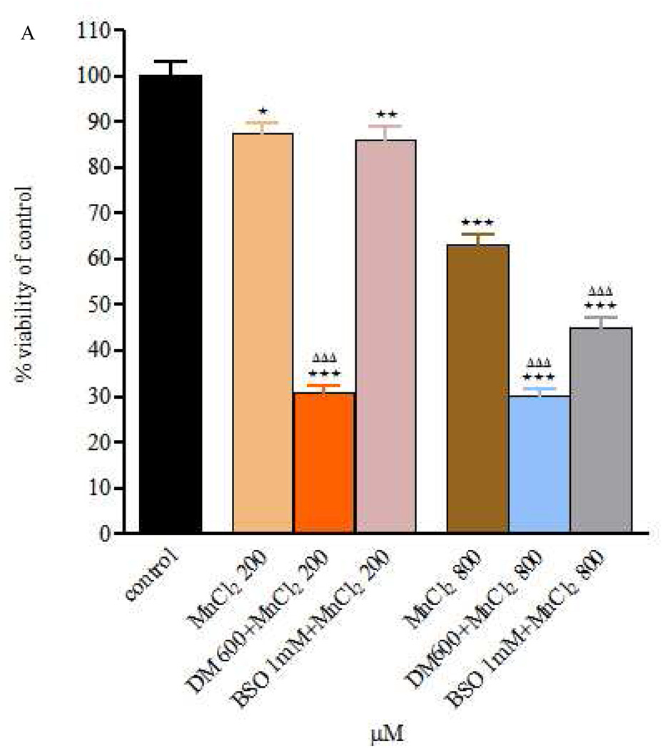
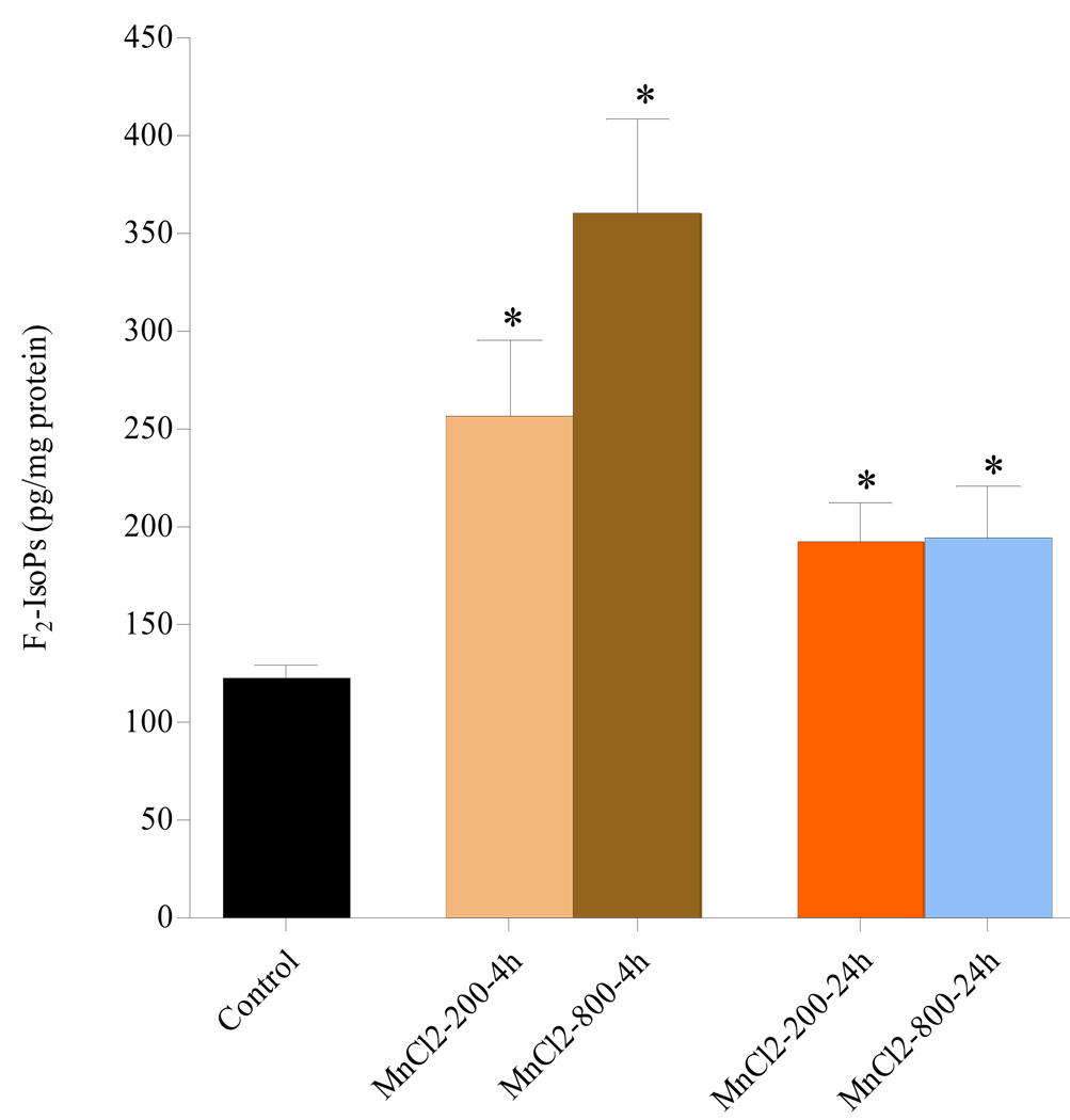
Manganese (Mn) is an essential trace metal; however, exposure to high Mn levels can result in neurodegenerative changes resembling Parkinson's disease (PD). Information on Mn's effects on endothelial cells of the blood-brain barrier (BBB) is lacking. Accordingly, we tested the hypothesis that BBB endothelial cells are a primary target for Mn-induced neurotoxicity. The studies were conducted in an in vitro BBB model of immortalized rat brain endothelial (RBE4) cells. ROS production was determined by F(2)-isoprostane (F(2)-IsoPs) measurement. The relationship between Mn toxicity and redox status was investigated upon intracellular glutathione (GSH) depletion with diethylmaleate (DEM) or L-buthionine sulfoximine (BSO). Mn exposure (200 or 800 microM MnCl(2) or MnSO(4)) for 4 or 24h led to significant decrease in cell viability vs. controls. DEM or BSO pre-treatment led to further enhancement in cytotoxicity vs. exposure to Mn alone, with more pronounced cell death after 24-h DEM pre-treatment. F(2)-IsoPs levels in cells exposed to MnCl(2) (200 or 800 microM) were significantly increased after 4h and remained elevated 24h after exposure compared with controls. Consistent with the effects on cell viability and F(2)-IsoPs, treatment with MnCl(2) (200 or 800 microM) was also associated with a significant decrease in membrane potential. This effect was more pronounced in cells exposed to DEM plus MnCl(2) vs. cells exposed to Mn alone. We conclude that Mn induces direct injury to mitochondria in RBE4 cells. The ensuing impairment in energy metabolism and redox status may modify the restrictive properties of the BBB compromising its function.

摘要

锰(Mn)是一种必需的微量元素;然而,暴露于高水平的锰会导致类似于帕金森病(PD)的神经退行性变化。关于锰对血脑屏障(BBB)内皮细胞的影响的信息是缺乏的。因此,我们检验了假设,即 BBB 内皮细胞是 Mn 诱导的神经毒性的主要靶标。这些研究是在体外 BBB 模型中进行的,使用永生大鼠脑内皮(RBE4)细胞。通过 F(2)-异前列腺素(F(2)-IsoPs)测量来确定 ROS 的产生。通过二乙基马来酸(DEM)或 L-丁硫氨酸亚砜(BSO)耗尽细胞内谷胱甘肽(GSH)来研究 Mn 毒性与氧化还原状态之间的关系。暴露于 Mn(200 或 800μM MnCl(2)或 MnSO(4))4 或 24h 导致细胞活力与对照相比显著降低。与单独暴露于 Mn 相比,DEM 或 BSO 预处理导致细胞毒性进一步增强,与 24h DEM 预处理后细胞死亡更为明显。暴露于 MnCl(2)(200 或 800μM)的细胞中 F(2)-IsoPs 水平在 4h 后显著增加,并在暴露后 24h 仍保持升高,与对照组相比。与细胞活力和 F(2)-IsoPs 的影响一致,用 MnCl(2)(200 或 800μM)处理也与膜电位显著降低相关。与单独暴露于 Mn 相比,暴露于 DEM 加 MnCl(2)的细胞中这种作用更为明显。我们得出结论,Mn 诱导 RBE4 细胞中线粒体的直接损伤。随之而来的能量代谢和氧化还原状态的损伤可能会改变 BBB 的限制特性,从而损害其功能。

https://cdn.ncbi.nlm.nih.gov/pmc/blobs/2b82/2849855/22a3aa2fbbff/nihms181338f4a.jpg
https://cdn.ncbi.nlm.nih.gov/pmc/blobs/2b82/2849855/ed1f3cd5495b/nihms181338f1a.jpg
https://cdn.ncbi.nlm.nih.gov/pmc/blobs/2b82/2849855/c17cafc90b5c/nihms181338f2a.jpg
https://cdn.ncbi.nlm.nih.gov/pmc/blobs/2b82/2849855/0174ee880482/nihms181338f3.jpg
https://cdn.ncbi.nlm.nih.gov/pmc/blobs/2b82/2849855/22a3aa2fbbff/nihms181338f4a.jpg

相似文献

1
Rat brain endothelial cells are a target of manganese toxicity.大鼠脑内皮细胞是锰毒性的靶细胞。
Brain Res. 2010 Apr 22;1326:152-61. doi: 10.1016/j.brainres.2010.02.016. Epub 2010 Feb 17.
2
Up-regulation of P-glycoprotein expression by glutathione depletion-induced oxidative stress in rat brain microvessel endothelial cells.谷胱甘肽耗竭诱导的氧化应激上调大鼠脑微血管内皮细胞中P-糖蛋白的表达
J Neurochem. 2006 Sep;98(5):1465-73. doi: 10.1111/j.1471-4159.2006.03993.x.
3
Antioxidants prevent the cytotoxicity of manganese in RBE4 cells.抗氧化剂可预防锰对RBE4细胞的细胞毒性。
Brain Res. 2008 Oct 21;1236:200-5. doi: 10.1016/j.brainres.2008.07.125. Epub 2008 Aug 12.
4
Metallothionein induction attenuates the effects of glutathione depletors in rat hepatocytes.金属硫蛋白的诱导可减轻谷胱甘肽耗竭剂对大鼠肝细胞的影响。
Toxicol Sci. 1999 Jun;49(2):297-305. doi: 10.1093/toxsci/49.2.297.
5
BNIP3 up-regulation and mitochondrial dysfunction in manganese-induced neurotoxicity.锰诱导神经毒性中BNIP3的上调与线粒体功能障碍
Neurotoxicology. 2009 May;30(3):414-22. doi: 10.1016/j.neuro.2009.02.012. Epub 2009 Feb 25.
6
Manganese-induced neurotoxicity is differentially enhanced by glutathione depletion in astrocytoma and neuroblastoma cells.在星形细胞瘤和神经母细胞瘤细胞中,谷胱甘肽耗竭会差异性地增强锰诱导的神经毒性。
Neurochem Res. 2006 Nov;31(11):1349-57. doi: 10.1007/s11064-006-9179-7. Epub 2006 Oct 20.
7
Role of reactive oxygen species in cis-dichlorodiammineplatinum-induced cytotoxicity on bladder cancer cells.活性氧在顺式二氯二氨铂诱导膀胱癌细胞细胞毒性中的作用
Br J Cancer. 1997;76(2):206-10. doi: 10.1038/bjc.1997.363.
8
Effective depletion of glutathione in rat striatum and substantia nigra by L-buthionine sulfoximine in combination with 2-cyclohexene-1-one.L-丁硫氨酸亚砜胺与2-环己烯-1-酮联合使用可有效耗尽大鼠纹状体和黑质中的谷胱甘肽。
Life Sci. 1990;47(25):2333-42. doi: 10.1016/0024-3205(90)90272-s.
9
Depletion of glutathione after gamma irradiation modifies survival.γ射线照射后谷胱甘肽的消耗会改变存活率。
Radiat Res. 1991 Mar;125(3):267-76.
10
Cellular glutathione depletion by diethyl maleate or buthionine sulfoximine: no effect of glutathione depletion on the oxygen enhancement ratio.用马来酸二乙酯或丁硫氨酸亚砜胺消耗细胞内谷胱甘肽:谷胱甘肽消耗对氧增强比无影响。
Radiat Res. 1983 Nov;96(2):422-8.

引用本文的文献

1
The impact of manganese on vascular endothelium.锰对血管内皮的影响。
Toxicol Res. 2024 Aug 13;40(4):501-517. doi: 10.1007/s43188-024-00260-1. eCollection 2024 Oct.
2
D-Ribose-L-Cysteine Improves Glutathione Levels, Neuronal and Mitochondrial Ultrastructural Damage, Caspase-3 and GFAP Expressions Following Manganese-Induced Neurotoxicity.D-核糖-L-半胱氨酸可改善锰诱导的神经毒性后谷胱甘肽水平、神经元和线粒体超微结构损伤、半胱天冬酶-3 和 GFAP 的表达。
Neurotox Res. 2021 Dec;39(6):1846-1858. doi: 10.1007/s12640-021-00404-3. Epub 2021 Sep 4.
3
Effects of Incubation of Human Brain Microvascular Endothelial Cells and Astrocytes with Pyridostigmine Bromide, DEET, or Permethrin in the Absence or Presence of Metal Salts.

本文引用的文献

1
Could mitochondrial dysfunction play a role in manganese toxicity?线粒体功能障碍是否在锰毒性中起作用?
Environ Toxicol Pharmacol. 1999 Mar;7(1):49-57. doi: 10.1016/s1382-6689(98)00054-4.
2
Antioxidants prevent the cytotoxicity of manganese in RBE4 cells.抗氧化剂可预防锰对RBE4细胞的细胞毒性。
Brain Res. 2008 Oct 21;1236:200-5. doi: 10.1016/j.brainres.2008.07.125. Epub 2008 Aug 12.
3
Manganese transport in eukaryotes: the role of DMT1.真核生物中的锰转运:二价金属离子转运体1的作用
在不存在或存在金属盐的情况下,将人脑血管内皮细胞和星形胶质细胞与溴吡斯的明、避蚊胺或氯菊酯孵育的影响。
Int J Environ Res Public Health. 2020 Nov 11;17(22):8336. doi: 10.3390/ijerph17228336.
4
The impact of manganese on neurotransmitter systems.锰对神经递质系统的影响。
J Trace Elem Med Biol. 2020 Sep;61:126554. doi: 10.1016/j.jtemb.2020.126554. Epub 2020 May 20.
5
Manganese Is Essential for Neuronal Health.锰对神经元健康至关重要。
Annu Rev Nutr. 2015;35:71-108. doi: 10.1146/annurev-nutr-071714-034419. Epub 2015 May 13.
6
Comparison between 5-aminosalicylic acid (5-ASA) and para-aminosalicylic acid (4-PAS) as potential protectors against Mn-induced neurotoxicity.5-氨基水杨酸(5-ASA)与对氨基水杨酸(4-PAS)对锰诱导神经毒性的潜在保护作用比较。
Biol Trace Elem Res. 2013 Apr;152(1):113-6. doi: 10.1007/s12011-012-9597-0. Epub 2013 Jan 15.
7
Effect of chronic glutathione deficiency on the behavioral phenotype of Gclm-/- knockout mice.慢性谷胱甘肽缺乏对 Gclm-/- 敲除小鼠行为表型的影响。
Neurotoxicol Teratol. 2012 Jul;34(4):450-7. doi: 10.1016/j.ntt.2012.04.009. Epub 2012 May 10.
8
Microarray genomic profile of mitochondrial and oxidant response in manganese chloride treated PC12 cells.氯化锰处理的 PC12 细胞中线粒体和氧化应激反应的基因芯片基因组图谱。
Neurotoxicology. 2012 Mar;33(2):162-8. doi: 10.1016/j.neuro.2012.01.001. Epub 2012 Jan 18.
9
Manganese induces oxidative stress, redox state unbalance and disrupts membrane bound ATPases on murine neuroblastoma cells in vitro: protective role of silymarin.锰诱导体外鼠神经母细胞瘤细胞氧化应激、氧化还原状态失衡和破坏膜结合 ATP 酶:水飞蓟素的保护作用。
Neurochem Res. 2011 Aug;36(8):1546-57. doi: 10.1007/s11064-011-0483-5. Epub 2011 Apr 30.
10
Prolactin is a peripheral marker of manganese neurotoxicity.催乳素是锰神经毒性的外周标志物。
Brain Res. 2011 Mar 25;1382:282-90. doi: 10.1016/j.brainres.2011.01.047. Epub 2011 Jan 22.
Neurotoxicology. 2008 Jul;29(4):569-76. doi: 10.1016/j.neuro.2008.04.022. Epub 2008 May 14.
4
Mitochondrial-dependent manganese neurotoxicity in rat primary astrocyte cultures.大鼠原代星形胶质细胞培养中依赖线粒体的锰神经毒性
Brain Res. 2008 Apr 8;1203:1-11. doi: 10.1016/j.brainres.2008.01.079. Epub 2008 Feb 11.
5
Oxidative stress and neurotoxicity.氧化应激与神经毒性。
Chem Res Toxicol. 2008 Jan;21(1):172-88. doi: 10.1021/tx700210j. Epub 2007 Dec 4.
6
Measurement of chronic oxidative and inflammatory stress by quantification of isoketal/levuglandin gamma-ketoaldehyde protein adducts using liquid chromatography tandem mass spectrometry.使用液相色谱串联质谱法通过定量异酮醛/左旋谷胱甘肽γ-酮醛蛋白加合物来测量慢性氧化和炎症应激。
Nat Protoc. 2007;2(9):2079-91. doi: 10.1038/nprot.2007.298.
7
Manganese induces oxidative impairment in cultured rat astrocytes.锰可诱导培养的大鼠星形胶质细胞发生氧化损伤。
Toxicol Sci. 2007 Jul;98(1):198-205. doi: 10.1093/toxsci/kfm095. Epub 2007 Apr 27.
8
Manganese-induced neurotoxicity is differentially enhanced by glutathione depletion in astrocytoma and neuroblastoma cells.在星形细胞瘤和神经母细胞瘤细胞中,谷胱甘肽耗竭会差异性地增强锰诱导的神经毒性。
Neurochem Res. 2006 Nov;31(11):1349-57. doi: 10.1007/s11064-006-9179-7. Epub 2006 Oct 20.
9
Speciation of manganese in cells and mitochondria: a search for the proximal cause of manganese neurotoxicity.细胞和线粒体中锰的形态:探寻锰神经毒性的近端原因。
Neurotoxicology. 2006 Sep;27(5):765-76. doi: 10.1016/j.neuro.2006.05.002. Epub 2006 May 7.
10
Piracetam improves mitochondrial dysfunction following oxidative stress.吡拉西坦可改善氧化应激后的线粒体功能障碍。
Br J Pharmacol. 2006 Jan;147(2):199-208. doi: 10.1038/sj.bjp.0706459.