• 文献检索
  • 文档翻译
  • 深度研究
  • 学术资讯
  • Suppr Zotero 插件Zotero 插件
  • 邀请有礼
  • 套餐&价格
  • 历史记录
应用&插件
Suppr Zotero 插件Zotero 插件浏览器插件Mac 客户端Windows 客户端微信小程序
定价
高级版会员购买积分包购买API积分包
服务
文献检索文档翻译深度研究API 文档MCP 服务
关于我们
关于 Suppr公司介绍联系我们用户协议隐私条款
关注我们

Suppr 超能文献

核心技术专利:CN118964589B侵权必究
粤ICP备2023148730 号-1Suppr @ 2026

文献检索

告别复杂PubMed语法,用中文像聊天一样搜索,搜遍4000万医学文献。AI智能推荐,让科研检索更轻松。

立即免费搜索

文件翻译

保留排版,准确专业,支持PDF/Word/PPT等文件格式,支持 12+语言互译。

免费翻译文档

深度研究

AI帮你快速写综述,25分钟生成高质量综述,智能提取关键信息,辅助科研写作。

立即免费体验

基于内源性蛋白质和肽的对比剂在高级别脑肿瘤的磁共振成像中的应用。

MR imaging of high-grade brain tumors using endogenous protein and peptide-based contrast.

机构信息

Department of Radiology, Zhujiang Hospital, Southern Medical University, Guangzhou, Guangdong, China.

出版信息

Neuroimage. 2010 Jun;51(2):616-22. doi: 10.1016/j.neuroimage.2010.02.050. Epub 2010 Feb 24.

DOI:10.1016/j.neuroimage.2010.02.050
PMID:20188197
原文链接:https://pmc.ncbi.nlm.nih.gov/articles/PMC2856810/
Abstract

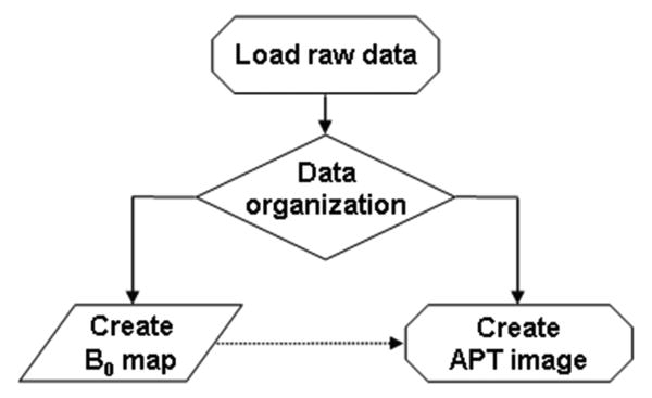
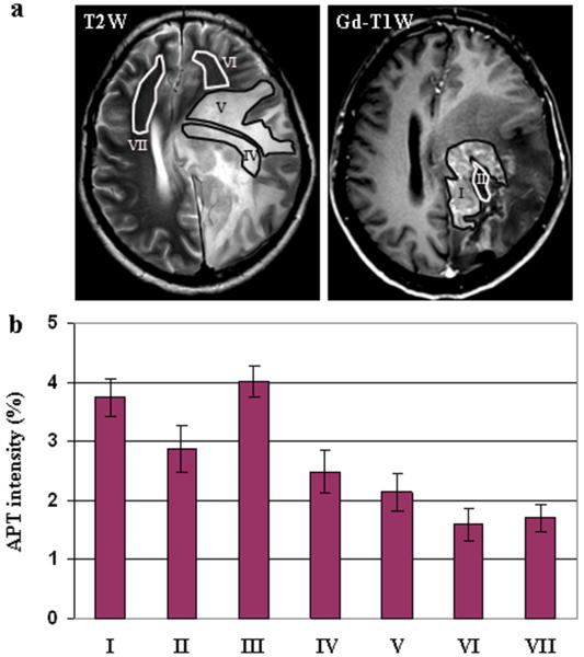
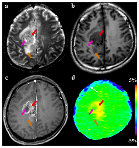
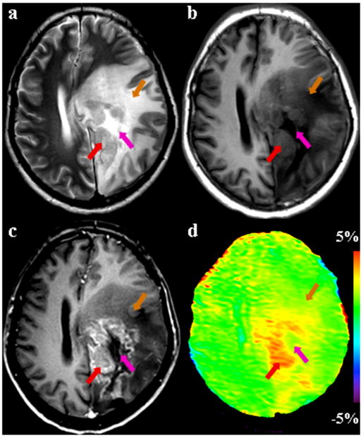
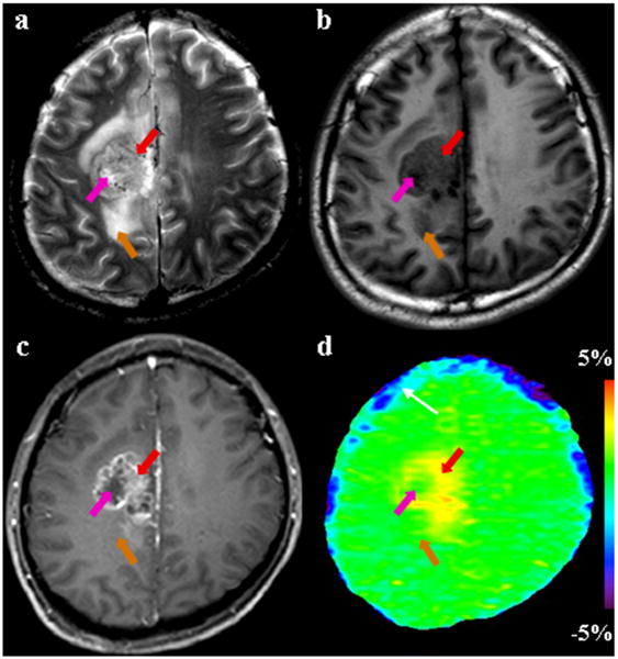
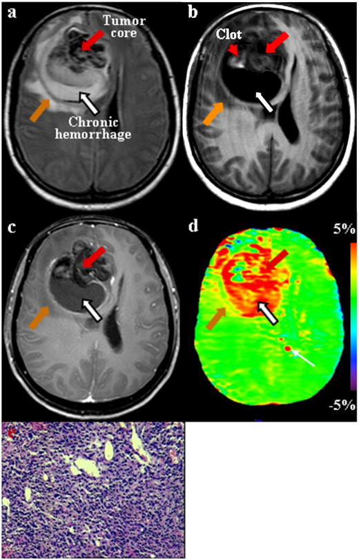
Amide proton transfer (APT) imaging is a novel MRI technique, in which the amide protons of endogenous proteins and peptides are irradiated to accomplish indirect detection using the bulk water signal. In this paper, the APT approach was added to a standard brain MRI protocol at 3T, and twelve patients with high-grade gliomas confirmed by histopathology were scanned. It is shown that all tumors, including one with minor gadolinium enhancement, showed heterogeneous hyperintensity on the APT images. The average APT signal intensities of the viable tumor cores were significantly higher than those of peritumoral edema and normal-appearing white matter (P<0.001). The average APT signal intensities were significantly lower in the necrotic regions than in the viable tumor cores (P=0.004). The APT signal intensities of the cystic cavities were similar to those of the viable tumor cores (P>0.2). The initial results show that APT imaging at the protein and peptide level may enhance non-invasive identification of tissue heterogeneity in high-grade brain tumors.

摘要

酰胺质子转移(APT)成像是一种新的 MRI 技术,利用该技术对内源性蛋白质和肽的酰胺质子进行照射,利用体纯水信号实现间接检测。在本文中,APT 方法被添加到标准的 3T 脑部 MRI 方案中,并对 12 名经组织病理学证实的高级别脑胶质瘤患者进行了扫描。结果显示,所有肿瘤,包括一个增强程度较小的肿瘤,在 APT 图像上均显示出不均匀的高信号。存活肿瘤核心的平均 APT 信号强度明显高于肿瘤周围水肿和正常白质(P<0.001)。坏死区的平均 APT 信号强度明显低于存活肿瘤核心(P=0.004)。囊性腔的 APT 信号强度与存活肿瘤核心相似(P>0.2)。初步结果表明,在蛋白质和肽水平上进行 APT 成像可能增强对高级别脑肿瘤组织异质性的无创识别。

https://cdn.ncbi.nlm.nih.gov/pmc/blobs/0c72/2856810/2d62721963aa/nihms183883f6.jpg
https://cdn.ncbi.nlm.nih.gov/pmc/blobs/0c72/2856810/fa82e64e61d8/nihms183883f1.jpg
https://cdn.ncbi.nlm.nih.gov/pmc/blobs/0c72/2856810/838115238950/nihms183883f2.jpg
https://cdn.ncbi.nlm.nih.gov/pmc/blobs/0c72/2856810/c1bb4d19b1bd/nihms183883f3.jpg
https://cdn.ncbi.nlm.nih.gov/pmc/blobs/0c72/2856810/b11e1747ef42/nihms183883f4.jpg
https://cdn.ncbi.nlm.nih.gov/pmc/blobs/0c72/2856810/58ad1daed98f/nihms183883f5.jpg
https://cdn.ncbi.nlm.nih.gov/pmc/blobs/0c72/2856810/2d62721963aa/nihms183883f6.jpg

相似文献

1
MR imaging of high-grade brain tumors using endogenous protein and peptide-based contrast.基于内源性蛋白质和肽的对比剂在高级别脑肿瘤的磁共振成像中的应用。
Neuroimage. 2010 Jun;51(2):616-22. doi: 10.1016/j.neuroimage.2010.02.050. Epub 2010 Feb 24.
2
Three-dimensional amide proton transfer MR imaging of gliomas: Initial experience and comparison with gadolinium enhancement.脑胶质瘤三维酰胺质子转移磁共振成像:初步经验及与钆增强对比
J Magn Reson Imaging. 2013 Nov;38(5):1119-28. doi: 10.1002/jmri.24067. Epub 2013 Feb 25.
3
Practical data acquisition method for human brain tumor amide proton transfer (APT) imaging.人脑肿瘤酰胺质子转移(APT)成像的实用数据采集方法。
Magn Reson Med. 2008 Oct;60(4):842-9. doi: 10.1002/mrm.21712.
4
Whole-brain amide proton transfer (APT) and nuclear overhauser enhancement (NOE) imaging in glioma patients using low-power steady-state pulsed chemical exchange saturation transfer (CEST) imaging at 7T.使用7T低功率稳态脉冲化学交换饱和转移(CEST)成像技术对胶质瘤患者进行全脑酰胺质子转移(APT)和核Overhauser增强(NOE)成像。
J Magn Reson Imaging. 2016 Jul;44(1):41-50. doi: 10.1002/jmri.25108. Epub 2015 Dec 10.
5
Amide proton transfer imaging of human brain tumors at 3T.3T下人脑肿瘤的酰胺质子转移成像
Magn Reson Med. 2006 Sep;56(3):585-92. doi: 10.1002/mrm.20989.
6
Grading diffuse gliomas without intense contrast enhancement by amide proton transfer MR imaging: comparisons with diffusion- and perfusion-weighted imaging.通过酰胺质子转移磁共振成像对无强化的弥漫性胶质瘤进行分级:与扩散加权成像和灌注加权成像的比较
Eur Radiol. 2017 Feb;27(2):578-588. doi: 10.1007/s00330-016-4328-0. Epub 2016 Mar 22.
7
APT-weighted and NOE-weighted image contrasts in glioma with different RF saturation powers based on magnetization transfer ratio asymmetry analyses.基于磁化转移率不对称分析的不同射频饱和功率下脑胶质瘤的 APT 加权和 NOE 加权图像对比。
Magn Reson Med. 2013 Aug;70(2):320-7. doi: 10.1002/mrm.24784. Epub 2013 May 9.
8
Evaluating the Role of Amide Proton Transfer (APT)-Weighted Contrast, Optimized for Normalization and Region of Interest Selection, in Differentiation of Neoplastic and Infective Mass Lesions on 3T MRI.评估酰胺质子转移(APT)加权对比在 3T MRI 中对肿瘤和感染性肿块病变的鉴别诊断中的作用,该对比针对标准化和感兴趣区选择进行了优化。
Mol Imaging Biol. 2020 Apr;22(2):384-396. doi: 10.1007/s11307-019-01382-x.
9
Quantitative assessment of amide proton transfer (APT) and nuclear overhauser enhancement (NOE) imaging with extrapolated semisolid magnetization transfer reference (EMR) signals: II. Comparison of three EMR models and application to human brain glioma at 3 Tesla.基于外推半固体磁化转移参考(EMR)信号的酰胺质子转移(APT)和核Overhauser增强(NOE)成像的定量评估:II. 三种EMR模型的比较及其在3特斯拉下对人脑胶质瘤的应用
Magn Reson Med. 2016 Apr;75(4):1630-9. doi: 10.1002/mrm.25795. Epub 2015 May 28.
10
Amide proton transfer imaging to discriminate between low- and high-grade gliomas: added value to apparent diffusion coefficient and relative cerebral blood volume.酰胺质子转移成像鉴别低级别与高级别胶质瘤:对表观扩散系数和相对脑血容量的附加价值
Eur Radiol. 2017 Aug;27(8):3181-3189. doi: 10.1007/s00330-017-4732-0. Epub 2017 Jan 23.

引用本文的文献

1
Double-Step R1rho-Based Lorentzian Fitting (DROF): A New CEST Analysis Approach and Its Comparison With Existing Methods.基于双步R1rho的洛伦兹拟合(DROF):一种新的CEST分析方法及其与现有方法的比较。
NMR Biomed. 2025 Aug;38(8):e70082. doi: 10.1002/nbm.70082.
2
Human liver CEST imaging at 7 T: Impact of shimming.7T场强下人体肝脏化学交换饱和转移成像:匀场的影响
Magn Reson Med. 2025 Oct;94(4):1604-1615. doi: 10.1002/mrm.30557. Epub 2025 May 24.
3
Use of amide proton transfer (APT) imaging in the differentiation of pediatric low-grade brain tumors from tumor-like brain lesions.

本文引用的文献

1
Magnetization transfer phenomenon in the human brain at 7 T.人脑在 7T 下的磁化传递现象。
Neuroimage. 2010 Jan 1;49(1):272-81. doi: 10.1016/j.neuroimage.2009.08.022. Epub 2009 Aug 14.
2
Integration of preoperative anatomic and metabolic physiologic imaging of newly diagnosed glioma.新诊断神经胶质瘤术前解剖与代谢生理成像的整合
J Neurooncol. 2009 May;92(3):401-15. doi: 10.1007/s11060-009-9845-0. Epub 2009 Apr 9.
3
Practical data acquisition method for human brain tumor amide proton transfer (APT) imaging.人脑肿瘤酰胺质子转移(APT)成像的实用数据采集方法。
酰胺质子转移(APT)成像在小儿低度脑肿瘤与脑肿瘤样病变鉴别中的应用。
Neuroradiology. 2025 Mar 21. doi: 10.1007/s00234-025-03582-5.
4
The Environmental Impact of Medical Imaging Agents and the Roadmap to Sustainable Medical Imaging.医学成像剂的环境影响与可持续医学成像路线图
Adv Sci (Weinh). 2025 Mar;12(9):e2404411. doi: 10.1002/advs.202404411. Epub 2025 Feb 4.
5
Amide proton transfer weighted MRI measurements yield consistent and repeatable results in patients with gliomas: a prospective test-retest study.酰胺质子转移加权磁共振成像测量在胶质瘤患者中产生一致且可重复的结果:一项前瞻性重测研究。
Eur Radiol. 2025 Jun;35(6):3367-3379. doi: 10.1007/s00330-024-11197-2. Epub 2024 Dec 18.
6
Multiparametric Chemical Exchange Saturation Transfer MRI Detects Metabolic Changes in Mild Cognitive Impairment Cases at 3.0 Tesla.多参数化学交换饱和转移磁共振成像在3.0特斯拉磁场下检测轻度认知障碍患者的代谢变化。
Neurochem Res. 2024 Dec 9;50(1):51. doi: 10.1007/s11064-024-04307-5.
7
The Role of Amide Proton Transfer (APT)-Weighted Imaging in Glioma: Assessment of Tumor Grading, Molecular Profile and Survival in Different Tumor Components.酰胺质子转移(APT)加权成像在胶质瘤中的作用:不同肿瘤成分的肿瘤分级、分子特征及生存评估
Cancers (Basel). 2024 Aug 29;16(17):3014. doi: 10.3390/cancers16173014.
8
Radiomic feature reliability of amide proton transfer-weighted MR images acquired with compressed sensing at 3T.3T下采用压缩感知采集的酰胺质子转移加权磁共振图像的影像组学特征可靠性
Int J Imaging Syst Technol. 2024 Mar;34(2). doi: 10.1002/ima.23027. Epub 2024 Jan 30.
9
Joint - Space Image Reconstruction and Data Fitting for Chemical Exchange Saturation Transfer Magnetic Resonance Imaging.化学交换饱和传递磁共振成像的联合空间图像重建和数据拟合。
Tomography. 2024 Jul 15;10(7):1123-1138. doi: 10.3390/tomography10070085.
10
A Combination of Amide Proton Transfer, Tumor Blood Flow, and Apparent Diffusion Coefficient Histogram Analysis Is Useful for Differentiating Malignant from Benign Intracranial Tumors in Young Patients: A Preliminary Study.酰胺质子转移、肿瘤血流及表观扩散系数直方图分析相结合有助于鉴别年轻患者颅内良恶性肿瘤:一项初步研究
Diagnostics (Basel). 2024 Jun 12;14(12):1236. doi: 10.3390/diagnostics14121236.
Magn Reson Med. 2008 Oct;60(4):842-9. doi: 10.1002/mrm.21712.
4
Multiparametric tissue characterization of brain neoplasms and their recurrence using pattern classification of MR images.利用磁共振图像的模式分类对脑肿瘤及其复发进行多参数组织特征分析。
Acad Radiol. 2008 Aug;15(8):966-77. doi: 10.1016/j.acra.2008.01.029.
5
Clinical features, mechanisms, and management of pseudoprogression in malignant gliomas.恶性胶质瘤假性进展的临床特征、机制及处理
Lancet Oncol. 2008 May;9(5):453-61. doi: 10.1016/S1470-2045(08)70125-6.
6
Nephrogenic systemic fibrosis associated with gadolinium based contrast agents: a summary of the medical literature reporting.与钆基造影剂相关的肾源性系统性纤维化:医学文献报道综述
Eur J Radiol. 2008 May;66(2):230-4. doi: 10.1016/j.ejrad.2008.02.011. Epub 2008 Mar 26.
7
Amide proton transfer imaging of human brain tumors at 3T.3T下人脑肿瘤的酰胺质子转移成像
Magn Reson Med. 2006 Sep;56(3):585-92. doi: 10.1002/mrm.20989.
8
Perfusion, diffusion and spectroscopy values in newly diagnosed cerebral gliomas.新诊断脑胶质瘤的灌注、扩散及波谱值
NMR Biomed. 2006 Jun;19(4):463-75. doi: 10.1002/nbm.1059.
9
Radiation necrosis versus glioma recurrence: conventional MR imaging clues to diagnosis.放射性坏死与胶质瘤复发:传统磁共振成像的诊断线索
AJNR Am J Neuroradiol. 2005 Sep;26(8):1967-72.
10
Quantitative apparent diffusion coefficients and T2 relaxation times in characterizing contrast enhancing brain tumors and regions of peritumoral edema.定量表观扩散系数和T2弛豫时间在鉴别强化脑肿瘤及瘤周水肿区域中的应用
J Magn Reson Imaging. 2005 Jun;21(6):701-8. doi: 10.1002/jmri.20335.