• 文献检索
  • 文档翻译
  • 深度研究
  • 学术资讯
  • Suppr Zotero 插件Zotero 插件
  • 邀请有礼
  • 套餐&价格
  • 历史记录
应用&插件
Suppr Zotero 插件Zotero 插件浏览器插件Mac 客户端Windows 客户端微信小程序
定价
高级版会员购买积分包购买API积分包
服务
文献检索文档翻译深度研究API 文档MCP 服务
关于我们
关于 Suppr公司介绍联系我们用户协议隐私条款
关注我们

Suppr 超能文献

核心技术专利:CN118964589B侵权必究
粤ICP备2023148730 号-1Suppr @ 2026

文献检索

告别复杂PubMed语法,用中文像聊天一样搜索,搜遍4000万医学文献。AI智能推荐,让科研检索更轻松。

立即免费搜索

文件翻译

保留排版,准确专业,支持PDF/Word/PPT等文件格式,支持 12+语言互译。

免费翻译文档

深度研究

AI帮你快速写综述,25分钟生成高质量综述,智能提取关键信息,辅助科研写作。

立即免费体验

频域光学相干断层扫描形态学特征与黄斑水肿造影渗漏模式的相关性。

Correlation between morphologic features on spectral-domain optical coherence tomography and angiographic leakage patterns in macular edema.

机构信息

Department of Ophthalmology, University of California San Diego, Shiley Eye Center, 0946, Joan and Irwin Jacobs Retina Center, 9415 Campus Point Drive, La Jolla, CA 92037, USA.

出版信息

Retina. 2010 Mar;30(3):383-9. doi: 10.1097/IAE.0b013e3181cd4803.

DOI:10.1097/IAE.0b013e3181cd4803
PMID:20216291
原文链接:https://pmc.ncbi.nlm.nih.gov/articles/PMC2870721/
Abstract

PURPOSE

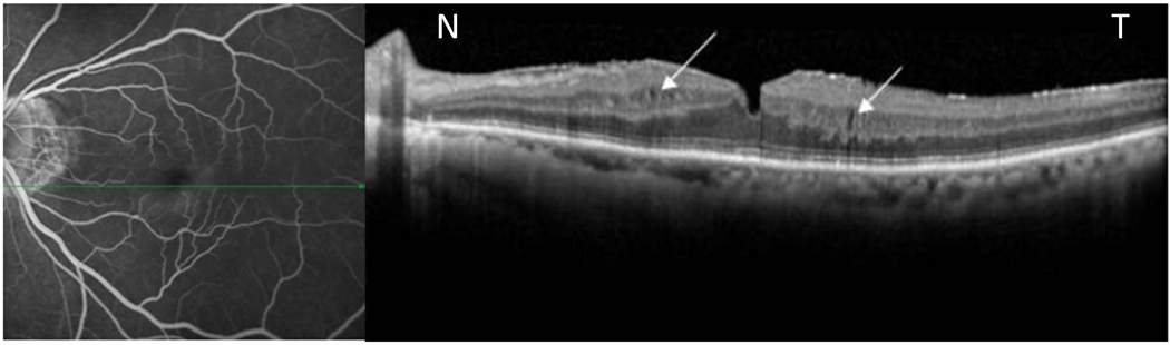
The purpose of this study was to determine the morphologic patterns of angiographic macular edema using simultaneous colocalization of fluorescein angiography and spectral-domain optical coherence tomography (SD-OCT) images in diabetes, epiretinal membrane, uveitic and pseudophakic cystoid macular edema, and vein occlusion.

METHODS

Eighty-seven consecutive patients (107 eyes) with macular edema from 5 different etiologies were imaged by simultaneous scanning laser ophthalmoscopy/OCT to study the morphologic patterns of edema on SD-OCT and then correlated/colocalized with the fluorescein angiographic patterns of leakage. Statistical analysis was done to analyze the differences in the morphologic OCT pattern by different diseases.

RESULTS

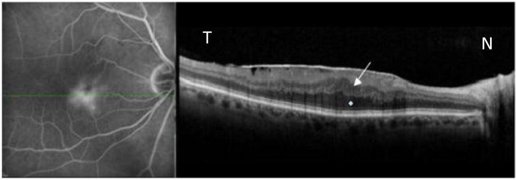
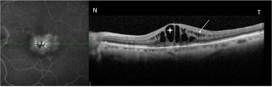
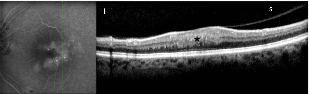
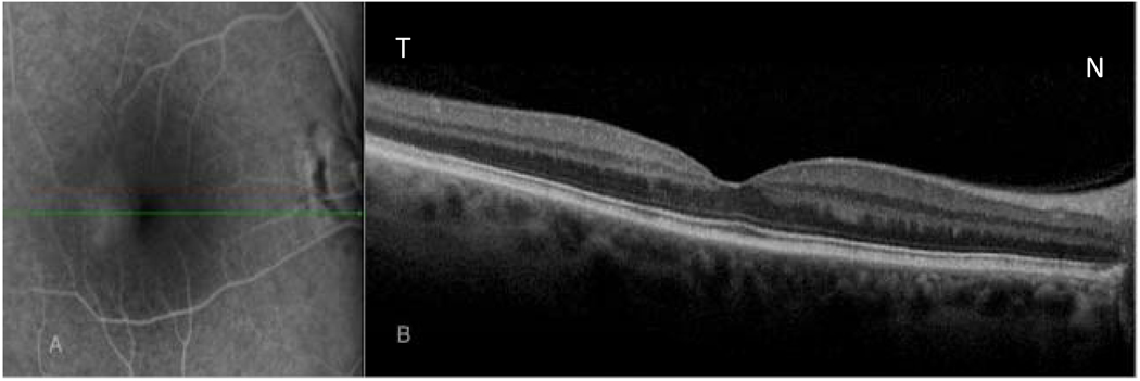
Spectral-domain OCT characteristics of macular edema showed a significant difference across different diseases (P = 0.037). Cystic fluid pockets were found to be more commonly seen in patients with diabetic macular edema and retinal vein occlusions, whereas those cases with macular edema secondary to epiretinal membrane showed noncystic changes on OCT. Seventy of the 107 eyes had diffuse angiographic leakage, and the remaining 37 eyes had cystoid leakage on angiography. Of the 70 eyes with diffuse leakage, 24.28% showed microcysts on SD-OCT in the area of edema, and 70% eyes had diffuse thickening or distorted architecture without cyst. All 37 eyes with cystoid leakage showed cysts in the area of edema by SD-OCT. A total of 3.73% of eyes with fluorescein angiographic leakage had no abnormalities on SD-OCT.

CONCLUSION

Eyes with diabetic macular edema and retinal vein occlusions have a significantly higher incidence of cyst formation on SD-OCT. There was no correlation between visual acuity and cyst formation. Diffuse noncystoid angiographic macular edema may show microcysts on SD-OCT, but diffuse edema is more commonly associated with thickening or distortion of the retinal layers without cyst formation. Cystoid leakage on fluorescein angiography is always associated with cystic changes on SD-OCT.

摘要

目的

本研究旨在通过共焦激光扫描检眼镜/光学相干断层扫描(OCT)同步扫描,确定糖尿病性、视网膜前膜性、葡萄膜炎性和后发性白内障性以及静脉阻塞性黄斑水肿的造影黄斑水肿的形态学模式。

方法

对 5 种不同病因引起黄斑水肿的 87 例连续患者(107 只眼)进行共焦激光扫描检眼镜/OCT 同步扫描,研究 OCT 上水肿的形态学模式,并与荧光素血管造影渗漏模式相关/共定位。通过不同疾病对 OCT 形态模式的差异进行统计学分析。

结果

不同疾病黄斑水肿的光谱 OCT 特征存在显著差异(P = 0.037)。在糖尿病性黄斑水肿和视网膜静脉阻塞患者中发现囊状液袋更为常见,而在由视网膜前膜引起的黄斑水肿患者中,OCT 显示为非囊状改变。107 只眼中 70 只眼有弥漫性造影渗漏,其余 37 只眼有囊样渗漏。在 70 只弥漫性渗漏眼中,24.28%的水肿区在 SD-OCT 上显示微囊,70%的眼有弥漫性增厚或无囊的扭曲结构。所有 37 只出现囊样渗漏的眼在 SD-OCT 上均显示水肿区有囊。在 SD-OCT 上,有 3.73%的荧光素血管造影渗漏眼无异常。

结论

糖尿病性黄斑水肿和视网膜静脉阻塞患者 SD-OCT 上囊形成的发生率明显较高。视力与囊形成无相关性。弥漫性非囊状造影黄斑水肿在 SD-OCT 上可能显示微囊,但弥漫性水肿更常见于无囊形成的视网膜层增厚或扭曲。荧光素血管造影上的囊样渗漏总是与 SD-OCT 上的囊状改变相关。

https://cdn.ncbi.nlm.nih.gov/pmc/blobs/f2e8/2870721/1e8e3b003929/nihms181090f6.jpg
https://cdn.ncbi.nlm.nih.gov/pmc/blobs/f2e8/2870721/3591f3e50125/nihms181090f1.jpg
https://cdn.ncbi.nlm.nih.gov/pmc/blobs/f2e8/2870721/e300343cd244/nihms181090f2.jpg
https://cdn.ncbi.nlm.nih.gov/pmc/blobs/f2e8/2870721/5e1cbbef691b/nihms181090f3.jpg
https://cdn.ncbi.nlm.nih.gov/pmc/blobs/f2e8/2870721/66b48da2051b/nihms181090f4.jpg
https://cdn.ncbi.nlm.nih.gov/pmc/blobs/f2e8/2870721/81322d1054c0/nihms181090f5.jpg
https://cdn.ncbi.nlm.nih.gov/pmc/blobs/f2e8/2870721/1e8e3b003929/nihms181090f6.jpg

相似文献

1
Correlation between morphologic features on spectral-domain optical coherence tomography and angiographic leakage patterns in macular edema.频域光学相干断层扫描形态学特征与黄斑水肿造影渗漏模式的相关性。
Retina. 2010 Mar;30(3):383-9. doi: 10.1097/IAE.0b013e3181cd4803.
2
Differentiation of Underlying Pathologies of Macular Edema Using Spectral Domain Optical Coherence Tomography (SD-OCT).利用谱域光学相干断层扫描(SD-OCT)对黄斑水肿的潜在病理进行鉴别。
Ocul Immunol Inflamm. 2019;27(3):474-483. doi: 10.1080/09273948.2019.1603313.
3
Correlation between spectral domain optical coherence tomography findings and fluorescein angiography patterns in diabetic macular edema.糖尿病性黄斑水肿的频域光学相干断层扫描结果与荧光素血管造影模式之间的相关性
Ophthalmology. 2009 Jun;116(6):1158-67. doi: 10.1016/j.ophtha.2008.12.063. Epub 2009 Apr 23.
4
Uveitic macular oedema: correlation between optical coherence tomography patterns with visual acuity and fluorescein angiography.葡萄膜炎性黄斑水肿:光学相干断层扫描模式与视力及荧光素血管造影之间的相关性
Br J Ophthalmol. 2008 Jul;92(7):922-7. doi: 10.1136/bjo.2007.136846.
5
The correlation between fluorescein angiographic and optical coherence tomographic features in clinically significant diabetic macular edema.临床显著性糖尿病黄斑水肿中荧光素血管造影与光学相干断层扫描特征之间的相关性
Am J Ophthalmol. 2004 Feb;137(2):313-22. doi: 10.1016/j.ajo.2003.09.016.
6
Correlation between fluorescein angiography and spectral-domain optical coherence tomography in the diagnosis of cystoid macular edema.荧光素血管造影与频域光相干断层扫描在诊断囊样黄斑水肿中的相关性。
Br J Ophthalmol. 2010 Sep;94(9):1197-200. doi: 10.1136/bjo.2009.170589. Epub 2009 Dec 3.
7
Characterization of macular edema from various etiologies by optical coherence tomography.通过光学相干断层扫描对各种病因引起的黄斑水肿进行特征描述。
Am J Ophthalmol. 2005 Aug;140(2):200-6. doi: 10.1016/j.ajo.2005.02.053.
8
SD-OCT pattern of retinal venous occlusion with cystoid macular edema treated with Ozurdex®.玻璃体内注射Ozurdex®治疗视网膜静脉阻塞合并黄斑囊样水肿的频域光学相干断层扫描图像特征
Eur J Ophthalmol. 2011 Sep-Oct;21(5):631-6. doi: 10.5301/EJO.2011.7428.
9
Patterns of macular edema in patients with uveitis: qualitative and quantitative assessment using optical coherence tomography.葡萄膜炎患者黄斑水肿的模式:使用光学相干断层扫描进行定性和定量评估
Ophthalmology. 2004 May;111(5):946-53. doi: 10.1016/j.ophtha.2003.08.037.
10
Spectral domain optical coherence tomography classification of diabetic macular edema: a new proposal to clinical practice.谱域光学相干断层扫描对糖尿病性黄斑水肿的分类:对临床实践的新建议。
Graefes Arch Clin Exp Ophthalmol. 2020 Jun;258(6):1165-1172. doi: 10.1007/s00417-020-04640-9. Epub 2020 Mar 9.

引用本文的文献

1
A Novel Effect of Microaneurysms and Retinal Cysts on Capillary Perfusion in Diabetic Macular Edema: A Multimodal Imaging Study.微动脉瘤和视网膜囊肿对糖尿病性黄斑水肿中毛细血管灌注的新影响:一项多模态成像研究
J Clin Med. 2025 Apr 25;14(9):2985. doi: 10.3390/jcm14092985.
2
Baseline macular structural and vascular changes as predictive markers of visual improvement in central retinal vein occlusion.基线黄斑结构和血管变化作为中心视网膜静脉阻塞视力改善的预测指标。
North Clin Istanb. 2025 Apr 24;12(2):222-230. doi: 10.14744/nci.2025.26932. eCollection 2025.
3
Visual function and disability are associated with microcystic macular edema, macular and peripapillary vessel density in patients with neuromyelitis optica spectrum disorder.

本文引用的文献

1
Normative data for macular thickness by high-definition spectral-domain optical coherence tomography (spectralis).高清光谱域光学相干断层扫描(Spectralis)测量黄斑厚度的正常参考值。
Am J Ophthalmol. 2009 Aug;148(2):266-71. doi: 10.1016/j.ajo.2009.03.006. Epub 2009 May 9.
2
Intravitreal bevacizumab (Avastin) for neovascular age-related macular degeneration using a variable frequency regimen in eyes with no previous treatment.在未经治疗的眼中采用可变频率方案,玻璃体内注射贝伐单抗(阿瓦斯汀)治疗新生血管性年龄相关性黄斑变性
Clin Exp Ophthalmol. 2008 Nov;36(8):748-55. doi: 10.1111/j.1442-9071.2008.01873.x.
3
A systematic correlation of angiography and high-resolution optical coherence tomography in diabetic macular edema.
视神经脊髓炎谱系障碍患者的视觉功能和残疾与微囊性黄斑水肿、黄斑及视乳头周围血管密度相关。
Front Neurol. 2022 Nov 14;13:1019959. doi: 10.3389/fneur.2022.1019959. eCollection 2022.
4
3D Reconstruction of a Unitary Posterior Eye by Converging Optically Corrected Optical Coherence and Magnetic Resonance Tomography Images via 3D CAD.通过 3D CAD 将经光学矫正的相干光断层扫描和磁共振断层扫描图像汇聚进行整体后眼部的 3D 重建。
Transl Vis Sci Technol. 2022 Jul 8;11(7):24. doi: 10.1167/tvst.11.7.24.
5
The RAP study, report 3: Discoloration of the macular region in patients with macular neovascularization type 3.RAP 研究,报告 3:3 型黄斑新生血管患者黄斑区变色。
Acta Ophthalmol. 2022 Feb;100(1):e270-e277. doi: 10.1111/aos.14866. Epub 2021 Apr 5.
6
Fluorescein Angiography Findings in Eyes With Lamellar Macular Hole and Epiretinal Membrane Foveoschisis.板层黄斑裂孔伴视网膜内界膜黄斑前膜裂孔的荧光素血管造影表现。
Invest Ophthalmol Vis Sci. 2021 Jan 4;62(1):34. doi: 10.1167/iovs.62.1.34.
7
Perioperative intraretinal fluid observed using optical coherence tomography in the epiretinal membrane.术中应用光学相干断层扫描观察眼内视网膜液在视网膜前膜中的表现。
BMC Ophthalmol. 2020 Jan 22;20(1):33. doi: 10.1186/s12886-019-1289-5.
8
2D Modeling and Correction of Fan-beam Scan Geometry in OCT.光学相干断层扫描中扇束扫描几何结构的二维建模与校正
Comput Pathol Ophthalmic Med Image Anal (2018). 2018 Sep;11039:328-335. doi: 10.1007/978-3-030-00949-6_39. Epub 2018 Sep 14.
9
Bimodal imaging of proliferative diabetic retinopathy vascular features using swept source optical coherence tomography angiography.使用扫频源光学相干断层扫描血管造影术对增殖性糖尿病视网膜病变血管特征进行双模态成像。
Int J Ophthalmol. 2018 Sep 18;11(9):1528-1533. doi: 10.18240/ijo.2018.09.16. eCollection 2018.
10
Optical coherence tomography: A guide to interpretation of common macular diseases.光学相干断层扫描:常见黄斑疾病解读指南
Indian J Ophthalmol. 2018 Jan;66(1):20-35. doi: 10.4103/ijo.IJO_902_17.
糖尿病性黄斑水肿中血管造影与高分辨率光学相干断层扫描的系统相关性研究
Ophthalmology. 2009 Jan;116(1):66-72. doi: 10.1016/j.ophtha.2008.09.042.
4
Uveitic macular oedema: correlation between optical coherence tomography patterns with visual acuity and fluorescein angiography.葡萄膜炎性黄斑水肿:光学相干断层扫描模式与视力及荧光素血管造影之间的相关性
Br J Ophthalmol. 2008 Jul;92(7):922-7. doi: 10.1136/bjo.2007.136846.
5
Discrepancy between fluorescein angiography and optical coherence tomography in detection of macular disease.荧光素血管造影与光学相干断层扫描在黄斑疾病检测中的差异
Retina. 2008 Apr;28(4):538-44. doi: 10.1097/IAE.0b013e318167270b.
6
Optical coherence tomography characteristics of group 2A idiopathic parafoveal telangiectasis.2A 组特发性黄斑旁毛细血管扩张症的光学相干断层扫描特征
Retina. 2007 Nov-Dec;27(9):1214-20. doi: 10.1097/IAE.0b013e318074bc4b.
7
Correlation between intraretinal changes in diabetic macular oedema seen in fluorescein angiography and optical coherence tomography.荧光素血管造影和光学相干断层扫描中所见糖尿病性黄斑水肿视网膜内变化的相关性
Acta Ophthalmol. 2008 Feb;86(1):34-9. doi: 10.1111/j.1600-0420.2007.00989.x. Epub 2007 Jul 25.
8
Intravitreal bevacizumab (Avastin) treatment of neovascular age-related macular degeneration.玻璃体内注射贝伐单抗(阿瓦斯汀)治疗新生血管性年龄相关性黄斑变性。
Retina. 2007 Apr-May;27(4):439-44. doi: 10.1097/IAE.0b013e31804b3e15.
9
An optical coherence tomography-guided, variable dosing regimen with intravitreal ranibizumab (Lucentis) for neovascular age-related macular degeneration.一项采用光学相干断层扫描引导的、玻璃体腔内注射雷珠单抗(Lucentis)可变给药方案治疗新生血管性年龄相关性黄斑变性的研究。
Am J Ophthalmol. 2007 Apr;143(4):566-83. doi: 10.1016/j.ajo.2007.01.028.
10
Correlation between optical coherence tomography and fluorescein angiography findings in diabetic macular edema.糖尿病性黄斑水肿中光学相干断层扫描与荧光素血管造影结果的相关性
Ophthalmology. 2007 Jan;114(1):104-7. doi: 10.1016/j.ophtha.2006.06.044. Epub 2006 Oct 27.