Costa Cássia Ax, Nunes Alvaro C, Ferreira Anderson J, Gomes Maria A, Caliari Marcelo V
Departamento de Patologia Geral, Instituto de Ciências Biológicas da Universidade Federal de Minas Gerais, Av, Antônio Carlos 6627, Belo Horizonte, Minas Gerais, Brasil.
Parasit Vectors. 2010 Mar 26;3(1):23. doi: 10.1186/1756-3305-3-23.
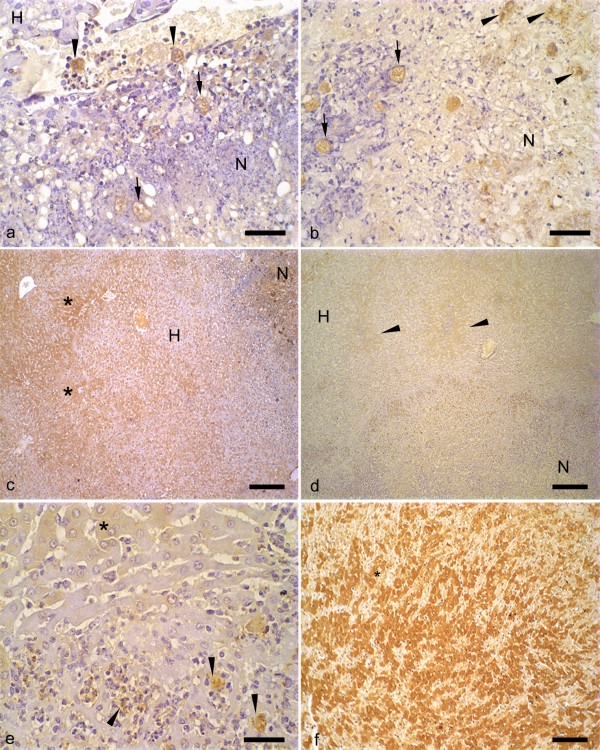
Human amoebiasis is caused by the parasitic protozoan Entamoeba histolytica that lives in the large intestine of hosts, where can produce asymptomatic colonization until severe invasive infections with blood diarrhea and spreading to other organs. The amoebic abscesses in liver are the most frequent form of amoebiasis outside intestine and still there are doubts about the pathogenic mechanisms involved in their formation. In this study we evaluated the in situ binding of antibodies, C3 and C9 complement components on trophozoites, in livers of hamsters infected with E. histolytica or E. dispar. These parameters were correlated with the extension of the hepatic lesions observed in these animals and with trophozoites survivor.
Hamsters were inoculated intra-hepatically with 100,000 trophozoites of E. histolytica or E. dispar strain and necropsied 12, 24, 48, 72, 144 and 192 h after inoculation. Antibodies, C3 and C9 binding to trophozoites were detected by immunohistochemistry. The estimation of the necrosis area and the number of labeled trophozoites was performed using digital morphometry analysis.
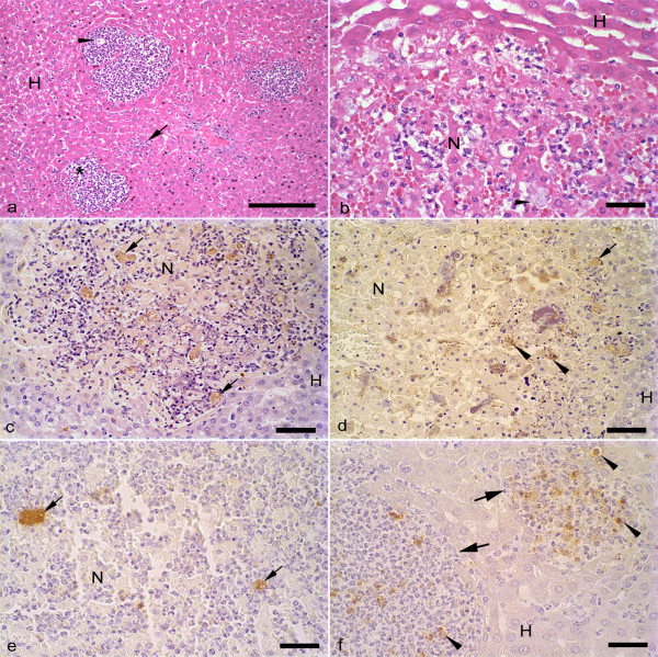
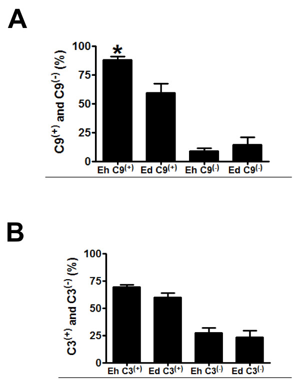
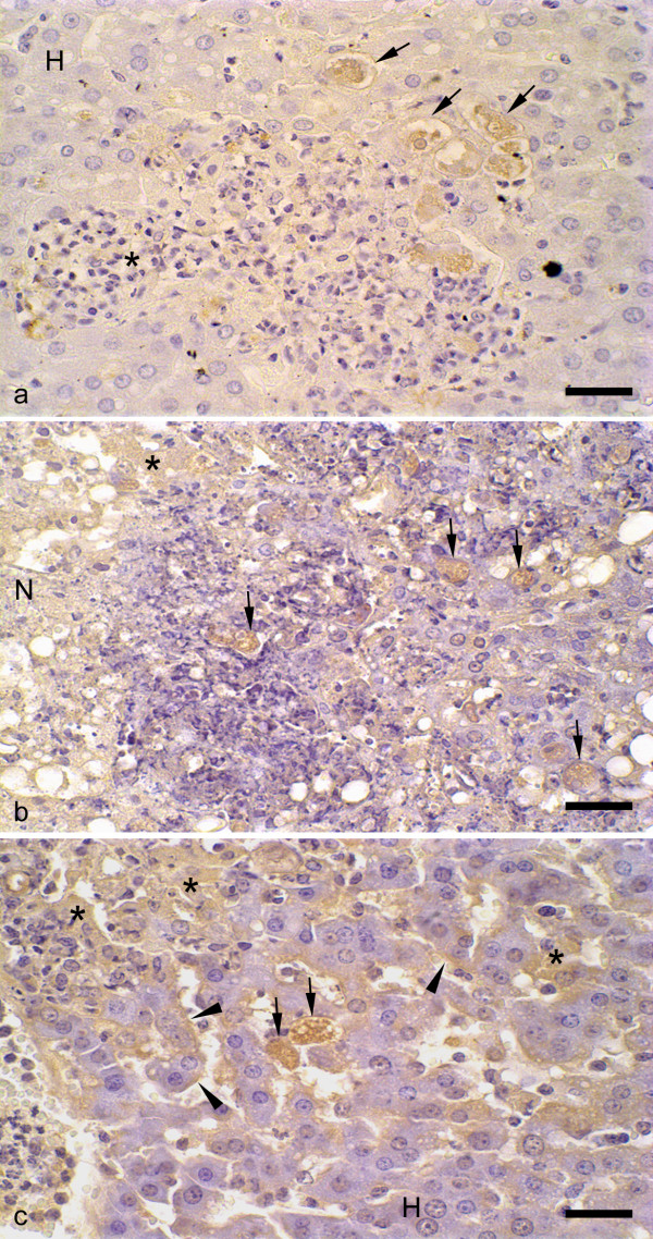
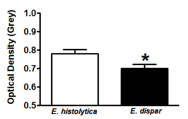
In the liver sections of animals inoculated with the amoebas, the binding of antibodies to E. histolytica trophozoites was significantly lower than to E. dispar trophozoites. Trophozoites of E. dispar were also more frequently vacuolated and high labeled cellular debris observed in the lesions. Positive diffuse reaction to C3 complement component was more intense in livers of animals inoculated with E. histolytica after 24 and 72 h of infection. C3(+) and C9(+) trophozoites were detected in the vascular lumen, granulomas and inside and in the border of necrotic areas of both infected group animals. C3(+) and C9(+) trophozoite debris immunostaining was higher in livers of E. dispar than in livers of E. histolytica. A positive correlation between necrotic areas and number of C9(+) trophozoites was observed in animals inoculated with E. dispar.
Morphological and immunohistochemical results suggest that antibodies and complement are able to bind and destroy some trophozoites in the liver of experimentally infected hamsters, perhaps selecting the more resistant parasites which are responsible by progression of amoebic abscesses. The findings indicate that E. histolytica possesses an enhanced ability in vivo to evade the immune responses compared to E. dispar, although it also causes experimental hepatic lesions.
人类阿米巴病由寄生原生动物溶组织内阿米巴引起,其寄生于宿主的大肠内,可导致无症状定植,直至发展为严重的侵袭性感染,出现血性腹泻并扩散至其他器官。肝阿米巴脓肿是肠道外最常见的阿米巴病形式,但其形成的致病机制仍存在疑问。在本研究中,我们评估了感染溶组织内阿米巴或迪斯帕内阿米巴的仓鼠肝脏中,抗体、C3和C9补体成分在滋养体上的原位结合情况。这些参数与这些动物肝脏病变的范围以及滋养体存活情况相关。
仓鼠经肝内接种100,000个溶组织内阿米巴或迪斯帕内阿米巴菌株的滋养体,并在接种后12、24、48、72、144和192小时进行剖检。通过免疫组织化学检测抗体、C3和C9与滋养体的结合情况。使用数字形态计量分析对坏死面积和标记滋养体的数量进行估计。
在接种阿米巴的动物肝脏切片中,抗体与溶组织内阿米巴滋养体的结合明显低于与迪斯帕内阿米巴滋养体的结合。迪斯帕内阿米巴的滋养体也更频繁地出现空泡化,并且在病变中观察到高标记的细胞碎片。感染后24小时和72小时,接种溶组织内阿米巴的动物肝脏中对C3补体成分的阳性弥漫反应更强。在两个感染组动物的血管腔、肉芽肿以及坏死区域内部和边界处均检测到C3(+)和C9(+)滋养体。迪斯帕内阿米巴感染组动物肝脏中C3(+)和C9(+)滋养体碎片的免疫染色高于溶组织内阿米巴感染组。在接种迪斯帕内阿米巴的动物中,观察到坏死区域与C9(+)滋养体数量之间呈正相关。
形态学和免疫组织化学结果表明,抗体和补体能够结合并破坏实验感染仓鼠肝脏中的一些滋养体,可能选择了导致阿米巴脓肿进展的更具抗性的寄生虫。研究结果表明,与迪斯帕内阿米巴相比,溶组织内阿米巴在体内具有更强的逃避免疫反应的能力,尽管它也会引起实验性肝脏病变。