Department of Biological Sciences, National University of Singapore, Singapore.
BMC Genomics. 2010 Mar 30;11:212. doi: 10.1186/1471-2164-11-212.
Mercury is a prominent environmental contaminant that causes detrimental effects to human health. Although the liver has been known to be a main target organ, there is limited information on in vivo molecular mechanism of mercury-induced toxicity in the liver. By using transcriptome analysis, phenotypic anchoring and validation of targeted gene expression in zebrafish, mercury-induced hepatotoxicity was investigated and a number of perturbed cellular processes were identified and compared with those captured in the in vitro human cell line studies.
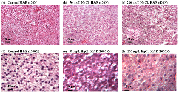
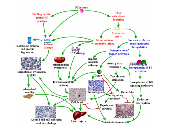
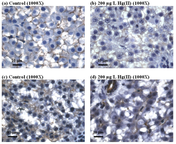
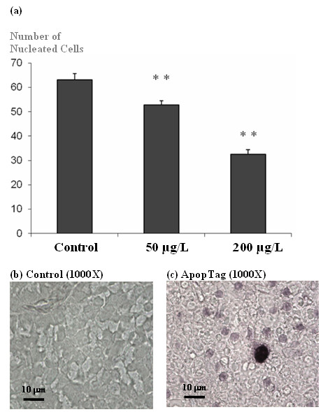
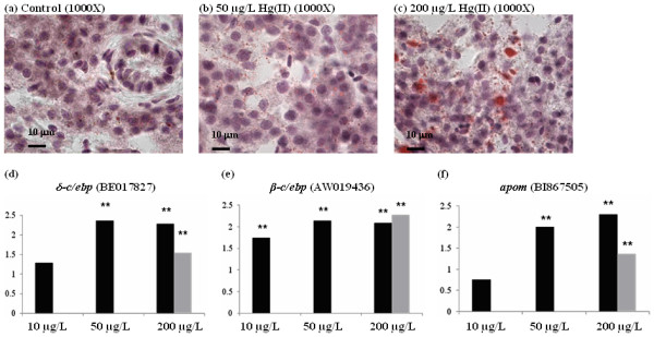
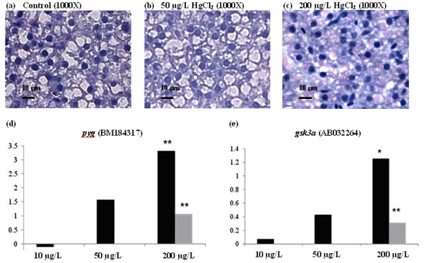
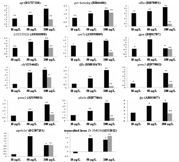
Hepato-transcriptome analysis of mercury-exposed zebrafish revealed that the earliest deregulated genes were associated with electron transport chain, mitochondrial fatty acid beta-oxidation, nuclear receptor signaling and apoptotic pathway, followed by complement system and proteasome pathway, and thereafter DNA damage, hypoxia, Wnt signaling, fatty acid synthesis, gluconeogenesis, cell cycle and motility. Comparative meta-analysis of microarray data between zebrafish liver and human HepG2 cells exposed to mercury identified some common toxicological effects of mercury-induced hepatotoxicity in both models. Histological analyses of liver from mercury-exposed fish revealed morphological changes of liver parenchyma, decreased nucleated cell count, increased lipid vesicles, glycogen and apoptotic bodies, thus providing phenotypic evidence for anchoring of the transcriptome analysis. Validation of targeted gene expression confirmed deregulated gene-pathways from enrichment analysis. Some of these genes responding to low concentrations of mercury may serve as toxicogenomic-based markers for detection and health risk assessment of environmental mercury contaminations.
Mercury-induced hepatotoxicity was triggered by oxidative stresses, intrinsic apoptotic pathway, deregulation of nuclear receptor and kinase activities including Gsk3 that deregulates Wnt signaling pathway, gluconeogenesis, and adipogenesis, leading to mitochondrial dysfunction, endocrine disruption and metabolic disorders. This study provides important mechanistic insights into mercury-induced liver toxicity in a whole-animal physiology context, which will help in understanding the syndromes caused by mercury poisoning. The molecular conservation of mercury-induced hepatotoxicity between zebrafish and human cell line reveals the feasibility of using zebrafish to model molecular toxicity in human for toxicant risk assessments.
汞是一种突出的环境污染物,对人类健康造成有害影响。尽管肝脏已被认为是主要的靶器官,但关于汞引起的肝毒性的体内分子机制的信息有限。通过使用转录组分析、表型锚定和靶向基因在斑马鱼中的表达验证,研究了汞诱导的肝毒性,并确定了一些受到干扰的细胞过程,并将其与体外人类细胞系研究中捕获的过程进行了比较。
暴露于汞的斑马鱼的肝转录组分析表明,最早失调的基因与电子传递链、线粒体脂肪酸β氧化、核受体信号和凋亡途径有关,其次是补体系统和蛋白酶体途径,然后是 DNA 损伤、缺氧、Wnt 信号、脂肪酸合成、糖异生、细胞周期和运动。暴露于汞的斑马鱼肝脏和人 HepG2 细胞的微阵列数据的比较荟萃分析确定了两种模型中汞诱导的肝毒性的一些共同毒性作用。暴露于汞的鱼肝脏的组织学分析显示肝实质的形态变化、核细胞计数减少、脂质泡、糖原和凋亡小体增加,从而为转录组分析的锚定提供了表型证据。靶向基因表达的验证证实了富集分析中失调的基因途径。这些对低浓度汞有反应的基因中的一些可能作为基于毒理学基因组的标志物,用于检测和评估环境汞污染的健康风险。
汞诱导的肝毒性是由氧化应激、内源性凋亡途径、核受体和激酶活性(包括调节 Wnt 信号通路、糖异生和脂肪生成的 Gsk3)的失调引起的,导致线粒体功能障碍、内分泌失调和代谢紊乱。这项研究为整体动物生理学背景下汞引起的肝毒性提供了重要的机制见解,有助于理解汞中毒引起的综合征。斑马鱼和人细胞系之间汞诱导肝毒性的分子保守性表明,使用斑马鱼对人类进行分子毒性建模进行毒物风险评估是可行的。