Crump Institute for Molecular Imaging, Department of Molecular and Medical Pharmacology, David Geffen School of Medicine, University of California Los Angeles, Los Angeles, CA 90095, USA.
Eur J Nucl Med Mol Imaging. 2010 Aug;37(8):1529-38. doi: 10.1007/s00259-010-1433-1. Epub 2010 Apr 1.
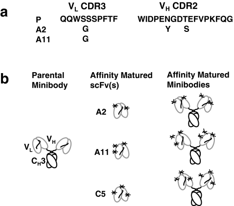
Prostate stem cell antigen (PSCA), a cell surface glycoprotein expressed in normal human prostate and bladder, is over-expressed in the majority of localized prostate cancer and most bone metastases. We have previously shown that the hu1G8 minibody, a humanized anti-PSCA antibody fragment (single-chain Fv-C(H)3 dimer, 80 kDa), can localize specifically and image PSCA-expressing xenografts at 21 h post-injection. However, the humanization and antibody fragment reformatting decreased its apparent affinity. Here, we sought to evaluate PET imaging contrast with affinity matured minibodies.
Yeast scFv display, involving four rounds of selection, was used to generate the three affinity matured antibody fragments (A2, A11, and C5) that were reformatted into minibodies. These three affinity matured anti-PSCA minibodies were characterized in vitro, and following radiolabeling with (124)I were evaluated in vivo for microPET imaging of PSCA-expressing tumors.
The A2, A11, and C5 minibody variants all demonstrated improved affinity compared to the parental (P) minibody and were ranked as follows: A2 > A11 > C5 > P. The (124)I-labeled A11 minibody demonstrated higher immunoreactivity than the parental minibody and also achieved the best microPET imaging contrast in two xenograft models, LAPC-9 (prostate cancer) and Capan-1 (pancreatic cancer), when evaluated in vivo.
Of the affinity variant minibodies tested, the A11 minibody that ranked second in affinity was selected as the best immunoPET tracer to image PSCA-expressing xenografts. This candidate is currently under development for evaluation in a pilot clinical imaging study.
前列腺干细胞抗原(PSCA)是一种在正常人类前列腺和膀胱中表达的细胞表面糖蛋白,在大多数局限性前列腺癌和大多数骨转移中过度表达。我们之前已经表明,hu1G8 迷你抗体,一种人源化抗 PSCA 抗体片段(单链 Fv-C(H)3 二聚体,80 kDa),可以在注射后 21 小时特异性定位并成像 PSCA 表达的异种移植物。然而,人源化和抗体片段的重新格式化降低了其表观亲和力。在这里,我们试图评估具有亲和力成熟的迷你抗体的 PET 成像对比。
酵母 scFv 展示,涉及四轮选择,用于生成三种亲和力成熟的抗体片段(A2、A11 和 C5),这些片段被重新格式化成立体。对这三种亲和力成熟的抗 PSCA 迷你抗体进行了体外表征,并在体内用(124)I 标记后,用于 PSCA 表达肿瘤的 microPET 成像评估。
A2、A11 和 C5 迷你抗体变体均显示出与亲本(P)迷你抗体相比具有改善的亲和力,并且按以下顺序排列:A2>A11>C5>P。(124)I 标记的 A11 迷你抗体比亲本迷你抗体具有更高的免疫反应性,并且在两个异种移植模型(LAPC-9[前列腺癌]和 Capan-1[胰腺癌])中进行体内评估时,也实现了最佳的 microPET 成像对比。
在所测试的亲和力变体迷你抗体中,排名第二的亲和力变体 A11 迷你抗体被选为成像 PSCA 表达异种移植物的最佳 immunoPET 示踪剂。该候选物目前正在开发中,用于评估一项初步的临床成像研究。