Scheper Ma, Chaisuparat R, Cullen Kj, Meiller Tf
Department of Oncology and Diagnostic Sciences and the Marlene and Stewart Greenebaum Cancer Center, University of Maryland Medical Center, 650 W Baltimore St, Baltimore, MD, 21201, USA.
Fibrogenesis Tissue Repair. 2010 Apr 1;3:6. doi: 10.1186/1755-1536-3-6.
Bisphosphonate (BP)-associated osteonecrosis of the jaw (ONJ) has been reported in patients receiving intravenous BP, particularly zoledronic acid (ZA). The purpose of this study was to develop an in vitro model representative of the effects BP has on soft tissue secondary to its release from bone. Human gingival fibroblasts and oral epithelial cell lines were exposed to various concentrations (0-10 muM) of ZA using dentine discs (DDs) as a direct carrier of BP, which were exposed for 24 hours to ZA in normal medium (NM), washed in phosphate-buffered saline (PBS) and placed in a new co-culture with the cells. The cells were allowed to proliferate until they grew over the bone discs and then the discs either were left unchelated, or were chelated using 0.001% EDTA or EGTA to release BP from the discs and to observe the cellular effects. Direct effects were determined using direct and fluorescent imaging. Apoptotic effects were determined by vital stain, terminal dUTP nick-end labeling, and annexin V studies. The effect on cell proliferation was determined by mitochondrial tetrazolium salt assay. The level of BP release was determined based on the effect of BP directly on cells, using the DDs or the supernatant fluids resulting from chelation.
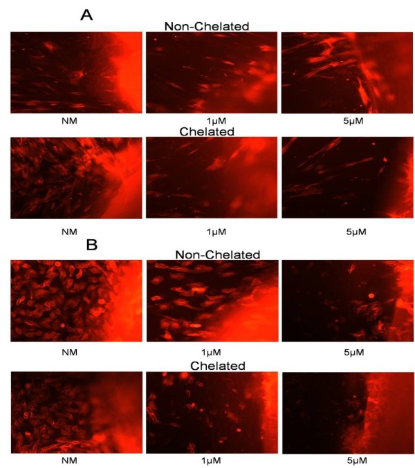
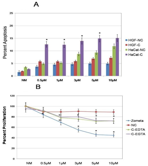
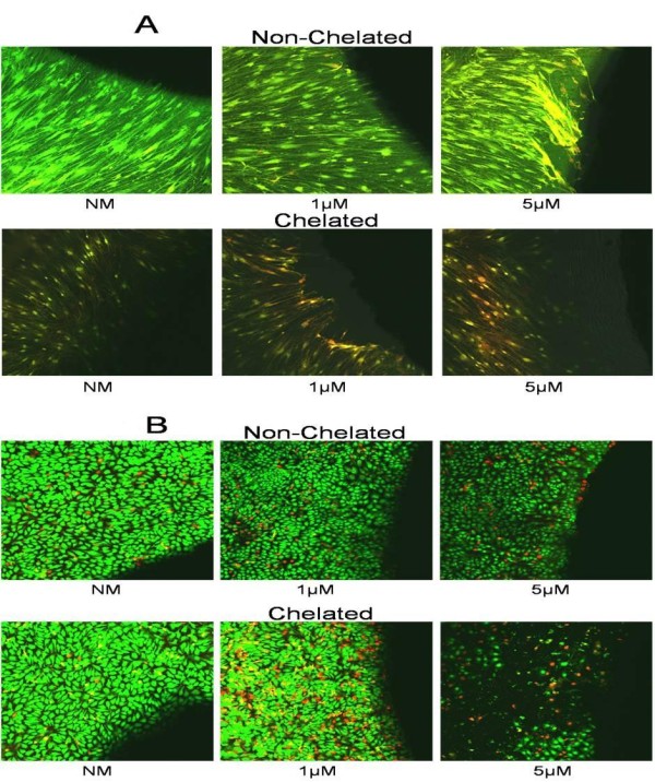
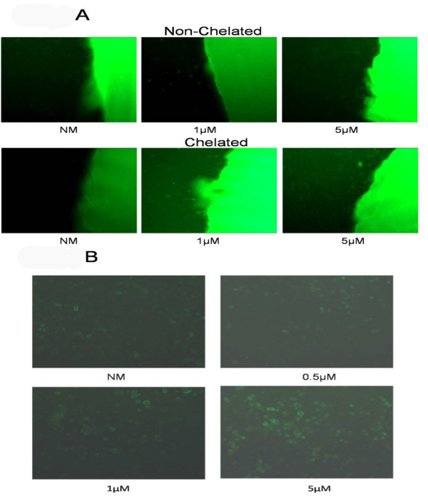
A dose-response effect was seen on imaging, and effects on apoptosis and cell proliferation were observed with increasing ZA concentrations liberated from the DDs, particularly after calcium cleavage and release of ZA from the DDs with a variety of chelating agents. Apoptotic effects were observed microscopically after chelation at 24 hours. Release of ZA was confirmed by extracting medium from non-chelated and chelated cell culture models with DDs and applying this medium to untreated fresh cell cultures, providing appropriate controls.
The results from this study demonstrate that low concentrations of ZA released from bone can rapidly and directly affect the oral mucosal tissues, initially through the induction of apoptosis and long term through the inhibition of cell proliferation. These findings provide an in vitro model for a soft-tissue mechanistic component in the initiation and/or progression of ONJ.
在接受静脉注射双膦酸盐(BP),尤其是唑来膦酸(ZA)的患者中,已报道了双膦酸盐相关的颌骨坏死(ONJ)。本研究的目的是建立一种体外模型,以代表BP从骨中释放后对软组织的影响。使用牙本质片(DDs)作为BP的直接载体,将人牙龈成纤维细胞和口腔上皮细胞系暴露于不同浓度(0 - 10 μM)的ZA中,牙本质片在正常培养基(NM)中暴露于ZA 24小时,用磷酸盐缓冲盐水(PBS)洗涤后,与细胞进行新的共培养。让细胞增殖,直到它们生长覆盖骨片,然后将骨片要么不进行螯合处理,要么用0.001%的乙二胺四乙酸(EDTA)或乙二醇双四乙酸(EGTA)进行螯合处理,以从骨片中释放BP并观察细胞效应。通过直接成像和荧光成像确定直接效应。通过活细胞染色、末端脱氧核苷酸转移酶介导的缺口末端标记法以及膜联蛋白V研究确定凋亡效应。通过线粒体四氮唑盐法确定对细胞增殖的影响。基于BP对细胞的直接作用,使用DDs或螯合产生的上清液来确定BP的释放水平。
在成像中观察到剂量反应效应,并且随着从DDs中释放的ZA浓度增加,观察到对凋亡和细胞增殖的影响,特别是在用各种螯合剂从DDs中进行钙裂解和释放ZA之后。在24小时螯合后通过显微镜观察到凋亡效应。通过从含有DDs的未螯合和螯合细胞培养模型中提取培养基,并将该培养基应用于未处理的新鲜细胞培养物(提供适当对照),证实了ZA的释放。
本研究结果表明,从骨中释放的低浓度ZA可迅速直接影响口腔黏膜组织,最初通过诱导凋亡,长期通过抑制细胞增殖。这些发现为ONJ起始和/或进展中的软组织机制成分提供了一种体外模型。