von Leithner Peter Lundh, Ciurtin Coziana, Jeffery Glen
Mol Vis. 2010 Mar 31;16:570-81.
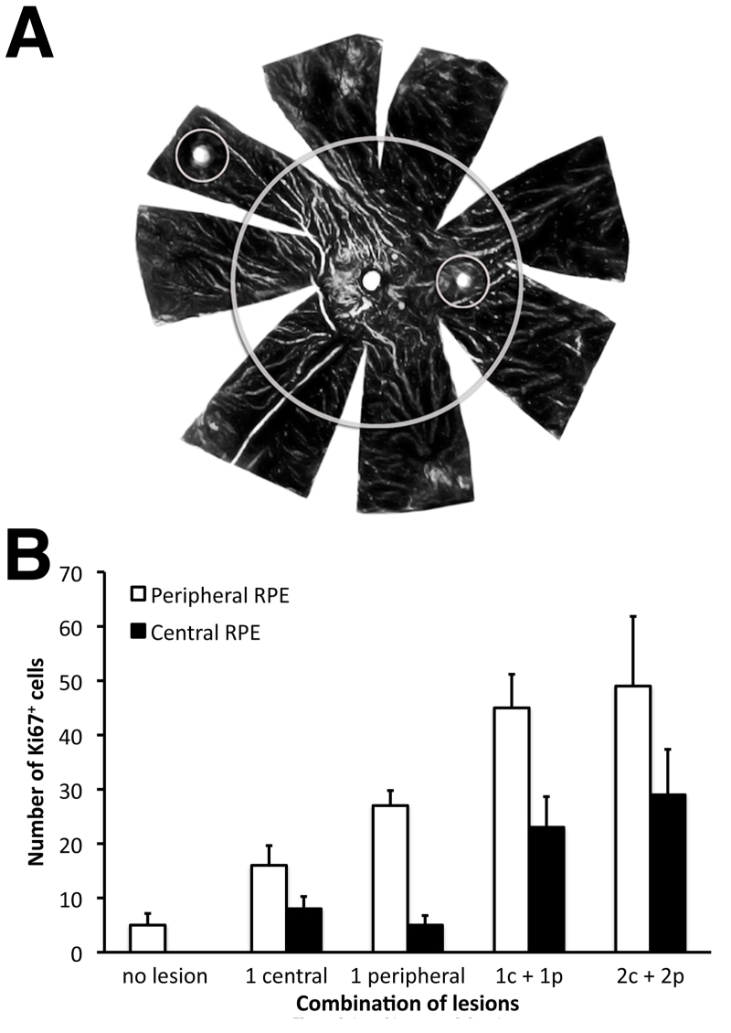
The vertebrate retina develops from the center to the periphery. In amphibians and fish the retinal margin continues to proliferate throughout life, resulting in retinal expansion. This does not happen in mammals. However, some mammalian peripheral retinal pigment epithelial (RPE) cells continue to divide, perhaps as a vestige of this mechanism. The RPE cells are adjacent to the ciliary margin, a known stem cell source. Here we test the hypothesis that peripheral RPE is fundamentally different from central RPE by challenging different regions with microscopic laser burns and charting differential responses in terms of levels of proliferation and the regions over which this proliferation occurs.
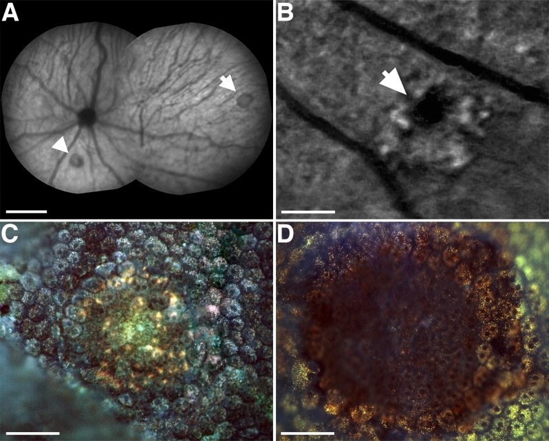
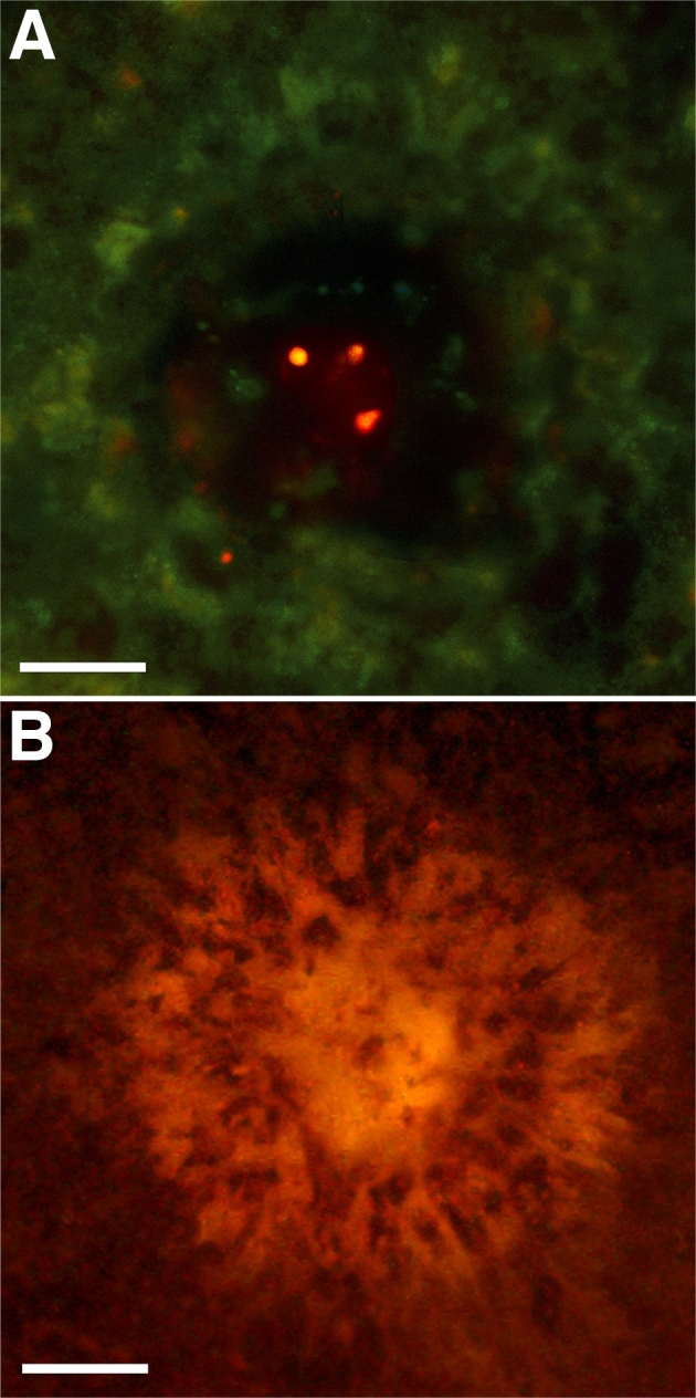
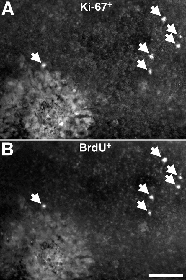
Microscopic RPE lesions were undertaken in rats at different eccentricities and the tissue stained for proliferative markers Ki67 and bromodeoxyuridine (BrdU) and the remodeling metalloproteinase marker 2 (MMP2).
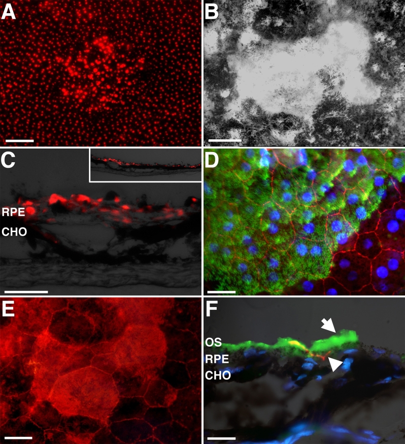
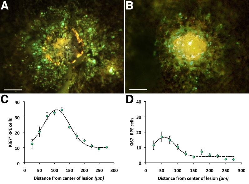
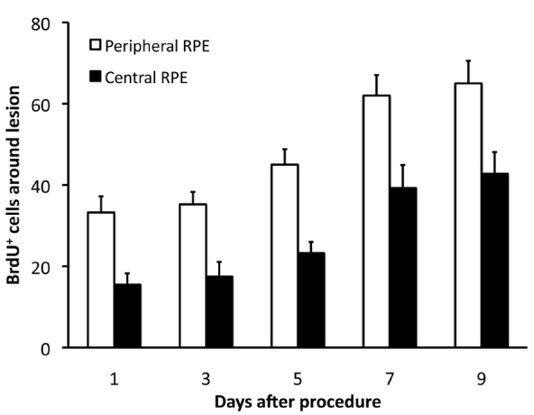
All lesions produced local RPE proliferation and tissue remodeling. Significantly more mitosis resulted from peripheral than central lesions. Unexpectedly, single lesions also resulted in RPE cells proliferating across the entire retina. Their number did not increase linearly with lesion number, indicating that they may be a specific population. All lesions repaired and formed apparently normal relations with the neural retina. Repaired RPE was albino.
These results highlight regional RPE differences, revealing an enhanced peripheral repair capacity. Further, all lesions have a marked impact on both local and distant RPE cells, demonstrating a pan retinal signaling mechanism triggering proliferation across the tissue plane. The RPE cells may represent a distinct population as their number did not increase with multiple lesions. The fact that repairing cells were hypopigmented is of interest because reduced pigment is associated with enhanced proliferative capacities in the developing neural retina.
脊椎动物的视网膜从中心向周边发育。在两栖动物和鱼类中,视网膜边缘在整个生命过程中持续增殖,导致视网膜扩张。哺乳动物中不会发生这种情况。然而,一些哺乳动物周边视网膜色素上皮(RPE)细胞会持续分裂,这可能是该机制的遗留现象。RPE细胞与睫状体边缘相邻,睫状体边缘是已知的干细胞来源。在此,我们通过用显微激光烧灼刺激不同区域,并绘制增殖水平和增殖发生区域方面的差异反应,来检验周边RPE与中央RPE存在根本差异这一假设。
在不同离心率的大鼠中进行显微RPE损伤,并对组织进行增殖标记物Ki67和溴脱氧尿苷(BrdU)以及重塑金属蛋白酶标记物2(MMP2)的染色。
所有损伤均导致局部RPE增殖和组织重塑。周边损伤产生的有丝分裂明显多于中央损伤。出乎意料的是,单个损伤也会导致RPE细胞在整个视网膜中增殖。它们的数量并不随损伤数量线性增加,这表明它们可能是一个特定的群体。所有损伤均得到修复,并与神经视网膜形成明显正常的关系。修复后的RPE呈白化状态。
这些结果突出了RPE的区域差异,揭示了周边修复能力增强。此外,所有损伤对局部和远处的RPE细胞都有显著影响,表明存在一种全视网膜信号机制,可触发跨组织平面的增殖。RPE细胞可能代表一个独特的群体,因为它们的数量不会随多个损伤而增加。修复细胞色素减退这一事实很有趣,因为色素减少与发育中的神经视网膜增殖能力增强有关。