Department of Ophthalmology, Emory University School of Medicine, Atlanta, Georgia, United States.
Department of Ophthalmology, Hallym University College of Medicine, Kangdong Sacred Heart Hospital, Seoul, South Korea.
Invest Ophthalmol Vis Sci. 2021 Feb 1;62(2):32. doi: 10.1167/iovs.62.2.32.
To quantitatively evaluate the changes in orientation and morphometric features of mouse retinal pigment epithelial (RPE) cells in different regions of the eye during aging.
We segmented individual RPE cells from whole RPE flatmount images of C57BL/6J mice (postnatal days 30 to 720) using a machine-learning method and evaluated changes in morphometric features, including our newly developed metric combining alignment and shape of RPE cells during aging.
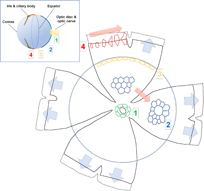
Mainly, the anterior part of the RPE sheet grows during aging, while the posterior part remains constant. Changes in size and shape of the peripheral RPE cells are prominent with aging as cells become larger, elongated, and concave. Conversely, the central RPE cells maintain relatively constant size and numbers with aging. Cell count in the central area and the overall cell count (approximately 50,000) were relatively constant over different age groups. RPE cells also present a specific orientation concordance that matches the shape of the specific region of the eyeball. Those cells near the optic disc or equator have a circumferential orientation to cover the round shape of the eyeball, whereas those cells in the periphery have a radial orientation and corresponding radial elongation, the extent of which increases with aging and matches with axial elongation of the eyeball.
These results suggest that the fluid RPE morphology reflects various growth rates of underlying eyeball, and RPE cells could be classified into four regional classes (near the optic disc, central, equatorial, and peripheral) according to their morphometric features.
定量评估小鼠视网膜色素上皮(RPE)细胞在衰老过程中眼睛不同区域的方向和形态特征的变化。
我们使用机器学习方法从 C57BL/6J 小鼠(出生后第 30 至 720 天)的整个 RPE 平片图像中分割单个 RPE 细胞,并评估形态特征的变化,包括我们新开发的结合 RPE 细胞对齐和形状的度量标准在衰老过程中的变化。
主要是 RPE 片的前部在衰老过程中生长,而后部保持不变。随着年龄的增长,周边 RPE 细胞的大小和形状变化明显,细胞变得更大、更长和更凹。相反,中央 RPE 细胞随着年龄的增长保持相对稳定的大小和数量。中央区域的细胞计数和总体细胞计数(约 50000 个)在不同年龄组中相对稳定。RPE 细胞还表现出特定的取向一致性,与眼球特定区域的形状相匹配。那些靠近视盘或赤道的细胞具有圆周取向以覆盖眼球的圆形,而那些在周边的细胞具有放射状取向和相应的放射状伸长,其程度随着年龄的增长而增加,并与眼球的轴向伸长相匹配。
这些结果表明,流体 RPE 形态反映了眼球下各种不同的生长速度,并且 RPE 细胞可以根据其形态特征分为四个区域类别(靠近视盘、中央、赤道和周边)。