Institute of Basic Medical Sciences, University of Oslo, Oslo, Norway.
Mol Biol Cell. 2010 Jun 1;21(11):1872-84. doi: 10.1091/mbc.e09-09-0839. Epub 2010 Apr 7.
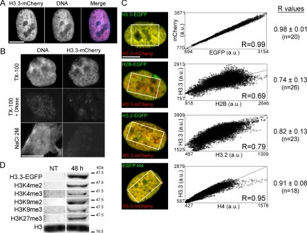
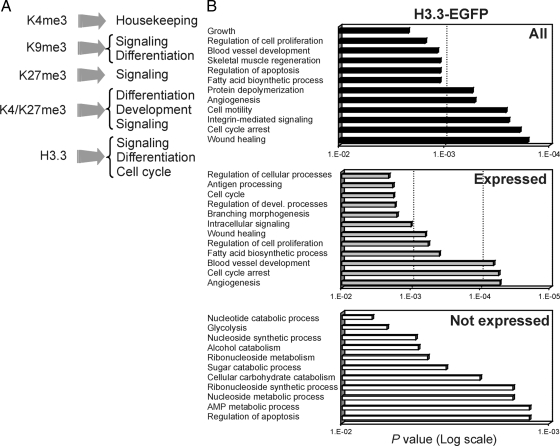
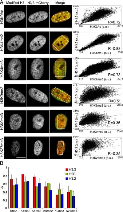
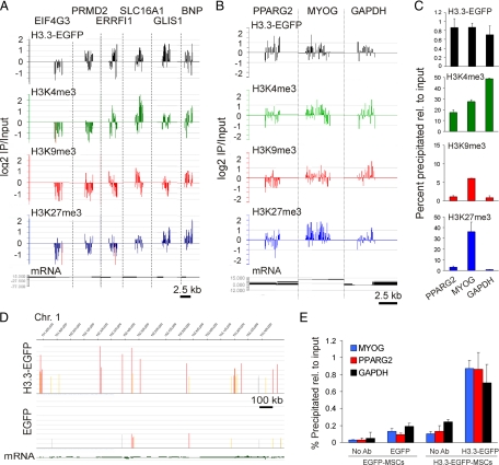
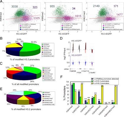
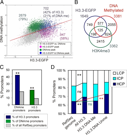
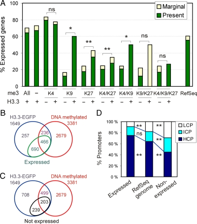
In contrast to canonical histones, histone variant H3.3 is incorporated into chromatin in a replication-independent manner. Posttranslational modifications of H3.3 have been identified; however, the epigenetic environment of incorporated H3.3 is unclear. We have investigated the genomic distribution of epitope-tagged H3.3 in relation to histone modifications, DNA methylation, and transcription in mesenchymal stem cells. Quantitative imaging at the nucleus level shows that H3.3, relative to replicative H3.2 or canonical H2B, is enriched in chromatin domains marked by histone modifications of active or potentially active genes. Chromatin immunoprecipitation of epitope-tagged H3.3 and array hybridization identified 1649 H3.3-enriched promoters, a fraction of which is coenriched in H3K4me3 alone or together with H3K27me3, whereas H3K9me3 is excluded, corroborating nucleus-level imaging data. H3.3-enriched promoters are predominantly CpG-rich and preferentially DNA methylated, relative to the proportion of methylated RefSeq promoters in the genome. Most but not all H3.3-enriched promoters are transcriptionally active, and coenrichment of H3.3 with repressive H3K27me3 correlates with an enhanced proportion of expressed genes carrying this mark. H3.3-target genes are enriched in mesodermal differentiation and signaling functions. Our data suggest that in mesenchymal stem cells, H3.3 targets lineage-priming genes with a potential for activation facilitated by H3K4me3 in facultative association with H3K27me3.
与经典组蛋白不同,组蛋白变体 H3.3 以复制独立的方式掺入染色质。已经鉴定了 H3.3 的翻译后修饰;然而,掺入的 H3.3 的表观遗传环境尚不清楚。我们研究了在间充质干细胞中,与组蛋白修饰、DNA 甲基化和转录相关的标记 H3.3 的基因组分布。核水平的定量成像显示,相对于复制的 H3.2 或经典的 H2B,H3.3 在染色质区域中富集,这些区域标记有活跃或潜在活跃基因的组蛋白修饰。标记的 H3.3 的染色质免疫沉淀和阵列杂交鉴定了 1649 个 H3.3 富集启动子,其中一部分与 H3K4me3 或 H3K27me3 共富集,而 H3K9me3 则被排除,这与核水平成像数据一致。H3.3 富集的启动子主要富含 CpG 且优先 DNA 甲基化,相对于基因组中甲基化 RefSeq 启动子的比例。大多数但不是所有的 H3.3 富集启动子都是转录活跃的,并且 H3.3 与抑制性 H3K27me3 的共富集与携带该标记的表达基因的比例增加相关。H3.3 靶基因在中胚层分化和信号转导功能中富集。我们的数据表明,在间充质干细胞中,H3.3 靶向具有激活潜力的谱系启动基因,这些基因通过 H3K4me3 与 H3K27me3 facultative 关联来促进激活。