Department of Pediatrics, Faculty of Medicine, Ain Shams University, Cairo, Egypt.
Diabetol Metab Syndr. 2010 Apr 15;2:23. doi: 10.1186/1758-5996-2-23.
Thalassemic patients suffer from diabetes mellitus secondary to hemosiderosis.
The study aimed to evaluate pancreatic iron overload by T2*-weighted Gradient-echo magnetic resonance imaging (MRI) in young beta-thalassemia major patients and to correlate it with glucose disturbances, hepatic hemosiderosis, serum ferritin and splenectomy.
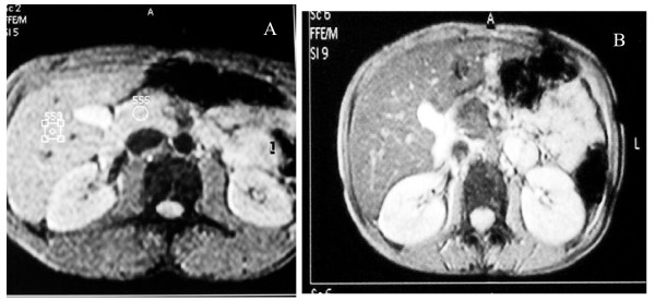
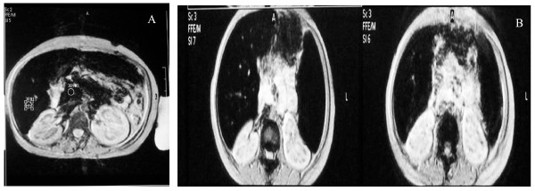
Forty thalassemic patients (20 non diabetic, 10 diabetic, and 10 with impaired glucose tolerance) were recruited from Pediatric Hematology Clinic, in addition to 20 healthy controls. All patients underwent clinical assessment and laboratory investigations included complete blood count, liver function tests, serum ferritin and oral glucose tolerance test (OGTT). A T2*-weighted gradient-echo sequence MRI was performed with 1.5 T scanner and signal intensity ratio (SIR) of the liver and the pancreas to noise were calculated.
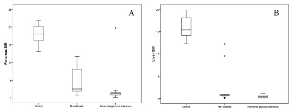
Significant reduction in signal intensity ratio (SIR) of the liver and the pancreas was shown in thalassemic patients compared to controls (P < 0.0001), Thalassemic patients with abnormal glucose tolerance; including diabetics and thalassemics with impaired glucose tolerance; displayed a higher degree of pancreatic and hepatic siderosis compared to thalassemics with normal glucose tolerance or controls (P < 0.001, P < 0.0001). Splenectomized thalassemic patients had significantly lower SIR of pancreas compared to non splenectomized patients (P < 0.05). A strong correlation was present between hepatic and pancreatic siderosis in studied patients (P < 0.001).
pancreatic siderosis can be detected by T2* gradient-echo MRI since childhood in thalassemic patients, and is more evident in patients with abnormal glucose tolerance. After splenectomy, iron deposition may be accelerated in the pancreas. Follow up of thalassemic patients using pancreatic MRI together with intensive chelation therapy may help to prevent the development of overt diabetes.
地中海贫血患者由于血色素沉着症而患有糖尿病。
本研究旨在通过 T2*-加权梯度回波磁共振成像(MRI)评估年轻β-地中海贫血主要患者的胰腺铁过载,并将其与葡萄糖紊乱、肝血色素沉着症、血清铁蛋白和脾切除术相关联。
从儿科血液学诊所招募了 40 名地中海贫血患者(20 名非糖尿病患者,10 名糖尿病患者和 10 名糖耐量受损患者),并招募了 20 名健康对照者。所有患者均接受了临床评估和实验室检查,包括全血细胞计数、肝功能检查、血清铁蛋白和口服葡萄糖耐量试验(OGTT)。使用 1.5 T 扫描仪进行 T2*-加权梯度回波序列 MRI,并计算肝脏和胰腺的信号强度比(SIR)。
与对照组相比,地中海贫血患者的肝脏和胰腺的信号强度比(SIR)明显降低(P <0.0001)。葡萄糖耐量异常的地中海贫血患者,包括糖尿病和糖耐量受损的地中海贫血患者,与葡萄糖耐量正常的地中海贫血患者或对照组相比,胰腺和肝铁沉着的程度更高(P <0.001,P <0.0001)。脾切除的地中海贫血患者的胰腺 SIR明显低于未脾切除的患者(P <0.05)。研究患者中存在肝和胰腺铁沉着之间的强烈相关性(P <0.001)。
T2*-梯度回波 MRI 可从小儿时期检测到地中海贫血患者的胰腺铁过载,并且在葡萄糖耐量异常的患者中更为明显。脾切除后,铁在胰腺中的沉积可能会加速。使用胰腺 MRI 对地中海贫血患者进行随访,并进行强化螯合治疗,可能有助于预防明显糖尿病的发生。