University College London Institute of Neurology, London, United Kingdom.
Biol Psychiatry. 2010 Jul 1;68(1):51-60. doi: 10.1016/j.biopsych.2010.03.019. Epub 2010 May 10.
Loss of cortical volume in frontotemporal regions has been reported in patients with schizophrenia and their relatives. Cortical area and thickness are determined by different genetic processes, and measuring these parameters separately may clarify disturbances in corticogenesis relevant to schizophrenia. Our study also explored clinical and cognitive correlates of these parameters.
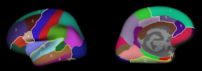
Thirty-seven patients with first-episode psychosis (34 schizophrenia, 3 schizoaffective disorder) and 38 healthy control subjects matched for age and sex took part in the study. Imaging was performed on an magnetic resonance imaging 1.5-T scanner. Area and thickness of the frontotemporal cortex were measured using a surface-based morphometry method (Freesurfer). All subjects underwent neuropsychologic testing that included measures of premorbid and current IQ, working and verbal memory, and executive function.
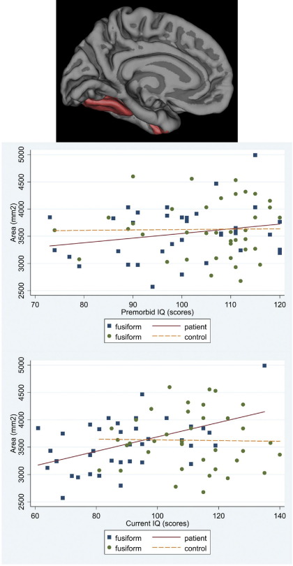
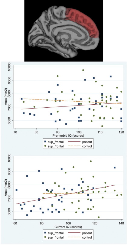
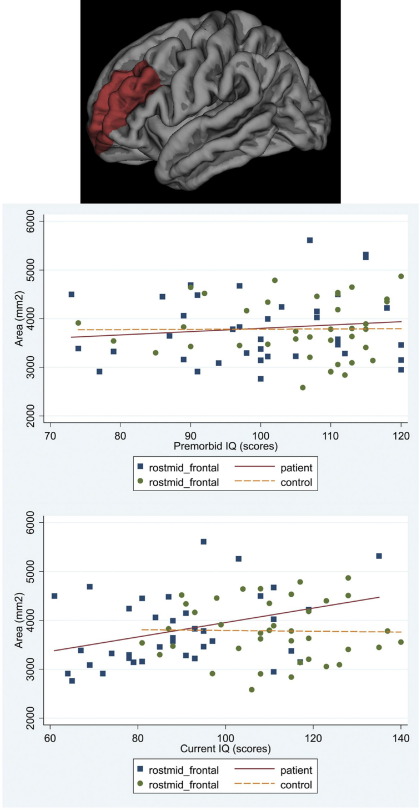
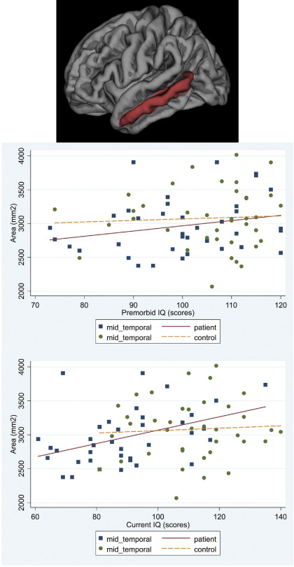
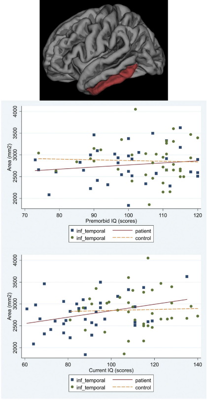
Reductions in cortical area, more marked in the temporal cortex, were present in patients. Overall frontotemporal cortical thickness did not differ between groups, although regional thinning of the right superior temporal region was observed in patients. There was a significant association of both premorbid IQ and IQ at disease onset with area, but not thickness, of the frontotemporal cortex, and working memory span was associated with area of the frontal cortex. These associations remained significant when only patients with schizophrenia were considered.
Our results suggest an early disruption of corticogenesis in schizophrenia, although the effect of subsequent environmental factors cannot be excluded. In addition, cortical abnormalities are subject to regional variations and differ from those present in neurodegenerative diseases.
额颞叶皮质体积的丧失已在精神分裂症患者及其亲属中报道。皮质面积和厚度由不同的遗传过程决定,分别测量这些参数可能会阐明与精神分裂症相关的皮质发生障碍。我们的研究还探讨了这些参数与临床和认知的相关性。
37 名首发精神病患者(34 名精神分裂症,3 名分裂情感障碍)和 38 名年龄和性别匹配的健康对照者参加了这项研究。成像在 1.5-T 磁共振成像扫描仪上进行。使用基于表面的形态计量学方法(Freesurfer)测量额颞皮质的面积和厚度。所有受试者均接受神经心理学测试,包括对既往和当前智商、工作记忆和言语记忆以及执行功能的测量。
患者存在皮质面积减少,颞叶皮质更明显。两组之间的额颞皮质总厚度没有差异,但患者右侧上颞叶区域存在局部变薄。无论是既往智商还是疾病发病时的智商,都与额颞皮质的面积显著相关,但与皮质厚度无关,工作记忆广度与额叶皮质的面积相关。当只考虑精神分裂症患者时,这些相关性仍然显著。
我们的结果表明精神分裂症存在皮质发生早期障碍,但不能排除随后环境因素的影响。此外,皮质异常存在区域性差异,与神经退行性疾病中的异常不同。