Department of Urology, Ludwig-Maximilians-University Munich, Marchionistr. 15, 81377 Munich, Germany.
Eur J Med Res. 2010 Mar 30;15(3):131-4. doi: 10.1186/2047-783x-15-3-131.
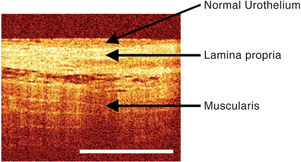
New modalities like Optical Coherence Tomography (OCT) allow non-invasive examination of the internal structure of biological tissue in vivo. The potential benefits and limitations of this new technology for the detection and evaluation of bladder cancer were examined in this study.
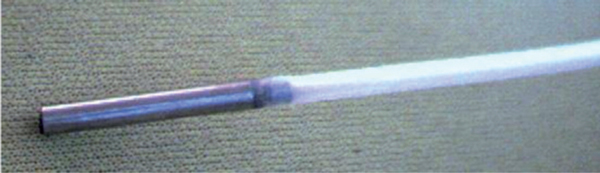
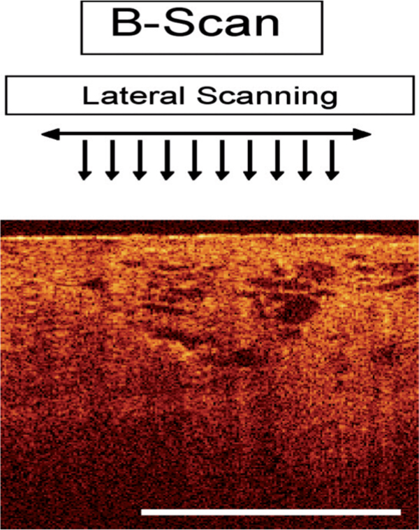
Between January 2007 and January 2008, 52 patients who underwent transurethral bladder biopsy or TUR-BT for surveillance or due to initial suspicion of urothelial carcinoma of the bladder were enrolled in this study. In total, 166 lesions were suspicious for malignancy according to standard white light cystoscopy. All suspicious lesions were scanned and interpreted during perioperative cystoscopy using OCT. Cold cup biopsies and/or TUR-B was performed for all these lesions. For this study we used an OCT-device (Niris, Imalux, Cleveland, US), that utilizes near-infrared light guided through a flexible fibre-based applicator, which is placed into the bladder via the working channel of the cystoscope. The technology provides high spatial resolution on the order of about 10-20 microm, and a visualization of tissue to a depth of about 2 mm across a lateral span of about 2 mm in width. The device used received market clearance from the FDA and CE approval in Germany. The diagnostic and surgical procedure was videotaped and analyzed afterwards for definitive matching of scanned and biopsied lesion. The primary aim of this study was to determine the level of correlation between OCT interpretation and final histological result.
Of 166 scanned OCT images, 102 lesions (61.4%) matched to the same site where the biopsy/TUR-BT was taken according to videoanalysis. Only these video-verified lesions were used for further analysis. Of all analyzed lesions 88 were benign (inflammation, edema, hyperplasia etc.) and 14 were malignant (CIS, Ta, T1, T2) as shown by final histo?pathology. - All 14 malignant lesions were detected correctly by OCT. Furthermore all invasive tumors were staged correctly by OCT regarding tumor growth beyond the lamina propria. There were no false negative lesions detected by OCT. Sensitivity of OCT for detecting the presence of a malignant lesion was 100% and sensitivity for detection of tumor growth beyond the lamina propria was 100% as well. Specificity of OCT for presence of malignancy was 65%, due to the fact that a number of lesions were interpreted as false positive by OCT.
As a minimally invasive technique, OCT proved to have extremely high sensitivity for detection of malignant lesions as well as estimation of whether a tumor has invaded beyond the lamina propria. However, specificity of OCT within the bladder was impaired (65%), possibly due to a learning curve and/or the relatively low spatial resolution and visualization depth of the OCT technology. Further studies and technical development are needed to establish an adequate surrogate for optical biopsy.
新的模式,如光学相干断层扫描(OCT),可以在体内无创地检查生物组织的内部结构。本研究旨在探讨这项新技术在膀胱癌的检测和评估中的潜在益处和局限性。
2007 年 1 月至 2008 年 1 月,对 52 例行经尿道膀胱活检或 TUR-BT 的患者进行了研究,这些患者行该检查的目的分别为监测或初始怀疑膀胱癌。根据标准的白光膀胱镜检查,共有 166 处病变疑似恶性。所有疑似病变均在围手术期膀胱镜检查中使用 OCT 进行扫描和解读。所有这些病变均进行冷杯活检和/或 TUR-B 检查。为了进行这项研究,我们使用了一种 OCT 设备(Niris,Imalux,克利夫兰,美国),该设备利用近红外光通过柔性光纤式探头引导,通过膀胱镜的工作通道进入膀胱。该技术提供了约 10-20μm 的高空间分辨率,以及约 2mm 深度的组织可视化,横向宽度约 2mm。该设备获得了 FDA 的市场批准和德国的 CE 认证。诊断和手术过程被录像,并在事后进行分析,以明确扫描和活检/ TUR-BT 病变的对应关系。本研究的主要目的是确定 OCT 解读与最终组织学结果之间的相关性水平。
在 166 张 OCT 扫描图像中,根据录像分析,有 102 处病变(61.4%)与活检/TUR-BT 的相同部位相匹配。仅对这些经录像验证的病变进行进一步分析。所有分析的病变中,88 处为良性(炎症、水肿、增生等),14 处为恶性(CIS、Ta、T1、T2),最终病理证实。所有 14 处恶性病变均被 OCT 正确检测到。此外,OCT 还正确地对肿瘤超出固有层的生长进行了分期。OCT 没有检测到假阴性病变。OCT 检测恶性病变的敏感性为 100%,检测肿瘤超出固有层的生长的敏感性也为 100%。OCT 检测恶性病变的特异性为 65%,因为一些病变被 OCT 错误地解读为假阳性。
作为一种微创技术,OCT 被证明对恶性病变的检测以及对肿瘤是否已超出固有层的评估具有极高的敏感性。然而,OCT 在膀胱内的特异性受损(65%),可能是由于学习曲线和/或 OCT 技术相对较低的空间分辨率和可视化深度。需要进一步的研究和技术发展,以建立一种合适的光学活检替代物。