Department of Morphological Sciences, School of Medicine-University Clinical Hospital, University of Santiago de Compostela, Santiago de Compostela, Spain.
Histopathology. 2010 Mar;56(4):472-80. doi: 10.1111/j.1365-2559.2010.03503.x.
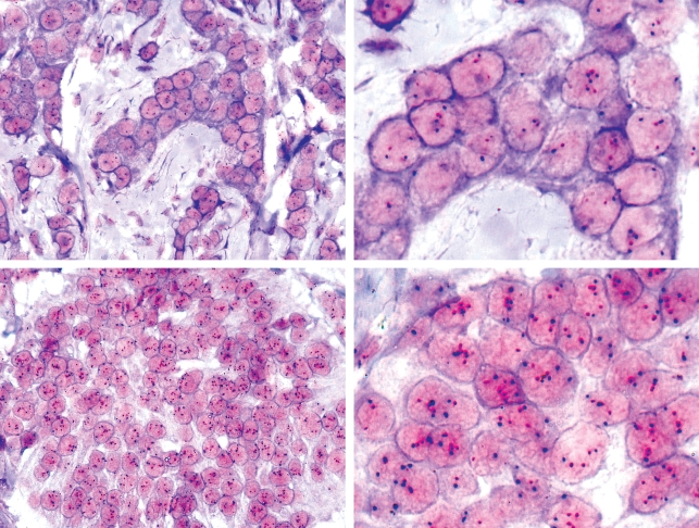
Fluorescence in situ hybridization (FISH) can be used to reveal several genomic imbalances relevant to proper cancer diagnosis and to the correct treatment regime. However, FISH requires expensive and advanced fluorescence microscopes in addition to expertise in fluorescence microscopy. To determine whether a newly developed dual-colour chromogenic in situ hybridization (CISH) method is a suitable alternative to FISH, we analysed the human epidermal growth factor receptor 2 gene (HER2) amplification level of 168 breast cancer specimens using dual-colour CISH and FISH and compared the results.
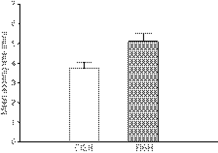
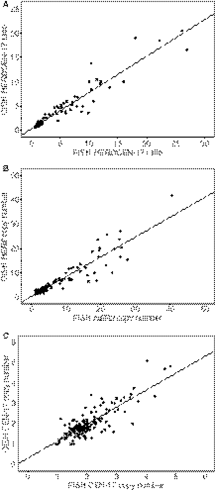
We found 100% agreement between HER2 status determined by FISH and dual-colour CISH. Furthermore, we observed that the time used to score slides was significantly reduced by 28% in dual-colour CISH compared with the FISH protocol. Concordance between HER2 protein status and dual-colour CISH or FISH was equally good with an overall agreement of 96.8%. Correlation between the HER2/centromere 17 gene ratios obtained with dual-colour CISH and FISH was highly significant with an overall correlation coefficient (rho) of 0.96.
We conclude that dual-colour CISH and bright field microscopy are excellent alternatives to FISH when analysing the HER2 status of primary breast cancer.
荧光原位杂交(FISH)可用于揭示与癌症正确诊断和正确治疗方案相关的几种基因组失衡。然而,FISH 需要昂贵且先进的荧光显微镜以及荧光显微镜方面的专业知识。为了确定新开发的双色显色原位杂交(CISH)方法是否是 FISH 的合适替代方法,我们使用双色 CISH 和 FISH 分析了 168 个乳腺癌标本的人表皮生长因子受体 2 基因(HER2)扩增水平,并比较了结果。
我们发现 FISH 确定的 HER2 状态与双色 CISH 之间有 100%的一致性。此外,我们观察到与 FISH 方案相比,双色 CISH 评分幻灯片的时间减少了 28%。HER2 蛋白状态与双色 CISH 或 FISH 的一致性同样良好,总一致性为 96.8%。双色 CISH 和 FISH 获得的 HER2/着丝粒 17 基因比值之间的相关性高度显著,总相关系数(rho)为 0.96。
我们得出结论,当分析原发性乳腺癌的 HER2 状态时,双色 CISH 和明场显微镜是 FISH 的极好替代方法。