Cissé Mamadou, Touré Alpha O, Konaté Ibrahima, Dieng Madieng, Ka Ousmane, Touré Fodé B, Dia Abdarahmane, Touré Cheikh T
Clinique Chirurgicale, Hôpital Aristide Le Dantec, Dakar, Avenue Pasteur, BP 3001, Sénégal.
J Med Case Rep. 2010 May 11;4:134. doi: 10.1186/1752-1947-4-134.
Situs inversus is a congenital anomaly characterized by the transposition of the abdominal viscera. When associated with dextrocardia, it is known as situs inversus totalis. This condition is rare and can be a diagnostic problem when associated with appendicular peritonitis.
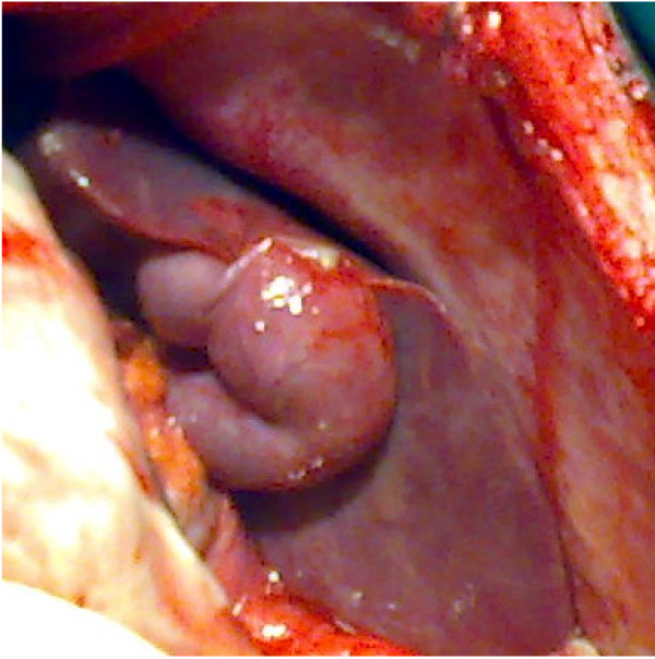
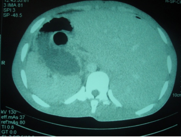
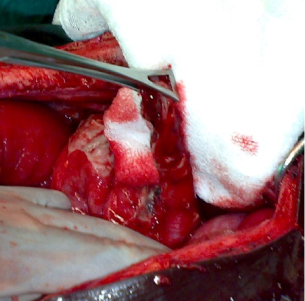
We report the case of a 20-year-old African man who presented to the emergency department with a 4-day history of diffuse abdominal pain, which began in his left iliac region and hypogastrium. After examination, we initiated a surgical exploration for peritonitis. We discovered a situs inversus at the left side of his liver, and his appendix was perforated in its middle third. A complementary post-operative thoracic and abdominal tomodensitometry revealed a situs inversus totalis.
Appendicular peritonitis in situs inversus is a rare association that can present a diagnostic problem. Morphologic exploration methods such as ultrasonography, tomodensitometry, magnetic resonance imaging, and laparoscopy may contribute to the early management of the disease and give guidance in choosing the most appropriate treatment for patients.
内脏反位是一种以腹腔脏器转位为特征的先天性异常。当与右位心相关时,称为完全性内脏反位。这种情况很罕见,与阑尾性腹膜炎相关时可能会成为诊断难题。
我们报告一例20岁非洲男性病例,该患者因弥漫性腹痛4天就诊于急诊科,腹痛始于左髂区和下腹部。检查后,我们因腹膜炎进行了手术探查。我们发现其肝脏位于左侧,存在内脏反位,且阑尾在其中段三分之一处穿孔。术后补充的胸部和腹部体层摄影术显示为完全性内脏反位。
内脏反位合并阑尾性腹膜炎是一种罕见的关联情况,可能会带来诊断问题。超声、体层摄影术、磁共振成像和腹腔镜检查等形态学探查方法可能有助于该疾病的早期处理,并为为患者选择最合适的治疗方法提供指导。