Omri S, Omri B, Savoldelli M, Jonet L, Thillaye-Goldenberg B, Thuret G, Gain P, Jeanny J C, Crisanti P, Behar-Cohen Francine
INSERM, U872 Physiopathology of ocular diseases: Therapeutic innovations, Paris, France.
Clin Ophthalmol. 2010 Apr 26;4:183-95. doi: 10.2147/opth.s5901.
The outer limiting membrane (OLM) is considered to play a role in maintaining the structure of the retina through mechanical strength. However, the observation of junction proteins located at the OLM and its barrier permeability properties may suggest that the OLM may be part of the retinal barrier.
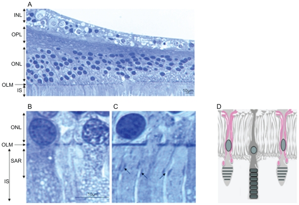
Normal and diabetic rat, monkey, and human retinas were used to analyze junction proteins at the OLM. Proteome analyses were performed using immunohistochemistry on sections and flat-mounted retinas and western blotting on protein extracts obtained from laser microdissection of the photoreceptor layers. Semi-thin and ultrastructure analyses were also reported.
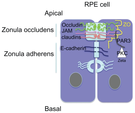
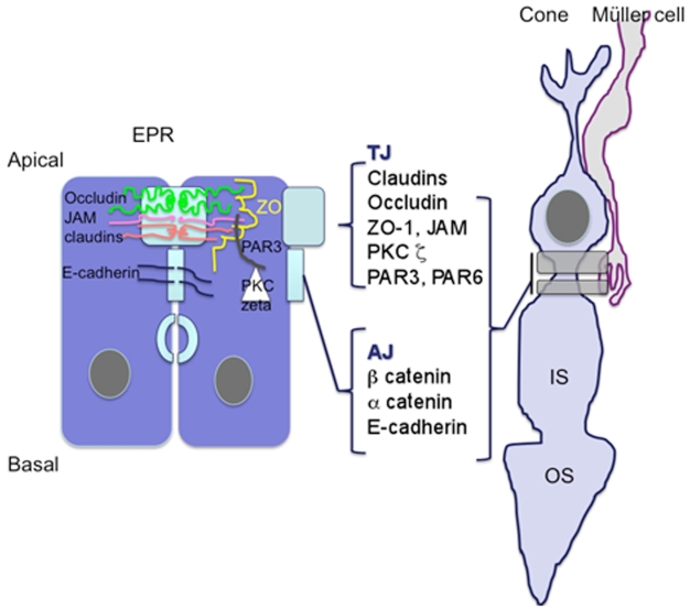
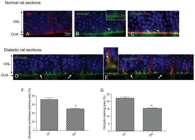
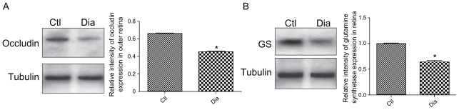
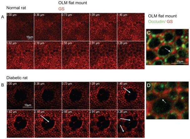
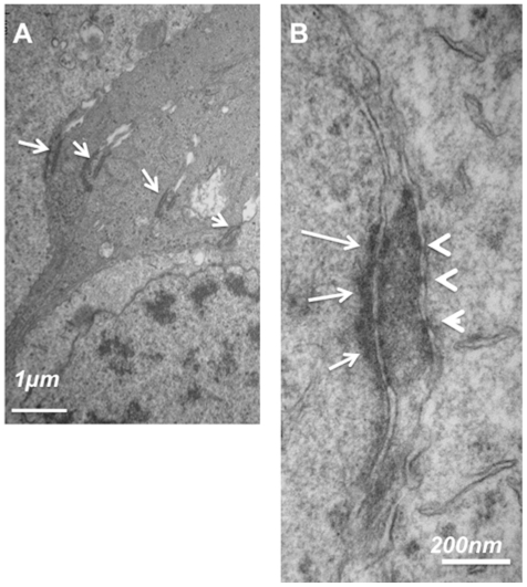
In the rat retina, in the subapical region zonula occludens-1 (ZO-1), junction adhesion molecule (JAM), an atypical protein kinase C, is present and the OLM shows dense labeling of occludin, JAM, and ZO-1. The presence of occludin has been confirmed using western blot analysis of the microdissected OLM region. In diabetic rats, occludin expression is decreased and glial cells junctions are dissociated. In the monkey retina, occludin, JAM, and ZO-1 are also found in the OLM. Junction proteins have a specific distribution around cone photoreceptors and Müller glia. Ultrastructural analyses suggest that structures like tight junctions may exist between retinal glial Müller cells and photoreceptors.
In the OLM, heterotypic junctions contain proteins from both adherent and tight junctions. Their structure suggests that tight junctions may exist in the OLM. Occludin is present in the OLM of the rat and monkey retina and it is decreased in diabetes. The OLM should be considered as part of the retinal barrier that can be disrupted in pathological conditions contributing to fluid accumulation in the macula.
外界限制膜(OLM)被认为通过机械强度在维持视网膜结构中发挥作用。然而,对位于OLM的连接蛋白及其屏障通透性特性的观察可能表明,OLM可能是视网膜屏障的一部分。
使用正常和糖尿病大鼠、猴及人类视网膜来分析OLM处的连接蛋白。采用免疫组织化学方法对切片和视网膜平铺标本进行蛋白质组分析,并对从光感受器层激光显微切割获得的蛋白质提取物进行蛋白质印迹分析。还报告了半薄切片和超微结构分析结果。
在大鼠视网膜中,在顶端下区域存在紧密连接蛋白1(ZO - 1)、连接黏附分子(JAM)、一种非典型蛋白激酶C,并且OLM显示出闭合蛋白、JAM和ZO - 1的密集标记。通过对显微切割的OLM区域进行蛋白质印迹分析证实了闭合蛋白的存在。在糖尿病大鼠中,闭合蛋白表达降低,神经胶质细胞连接解离。在猴视网膜中,也在OLM中发现了闭合蛋白、JAM和ZO - 1。连接蛋白在视锥光感受器和Müller神经胶质细胞周围有特定分布。超微结构分析表明,视网膜神经胶质Müller细胞和光感受器之间可能存在紧密连接样结构。
在OLM中,异型连接包含来自黏附连接和紧密连接的蛋白质。它们的结构表明OLM中可能存在紧密连接。闭合蛋白存在于大鼠和猴视网膜的OLM中,并且在糖尿病状态下减少。OLM应被视为视网膜屏障的一部分,在导致黄斑区积液的病理状况下可能会被破坏。