University of California, San Francisco, UCSF Box 0625, San Francisco, CA 94143-0625,
Osteoporos Int. 2011 Jan;22(1):85-90. doi: 10.1007/s00198-010-1265-7. Epub 2010 May 18.
While many assume hyperkyphosis reflects underlying spinal osteoporosis and vertebral fractures, our results suggest hyperkyphosis is independently associated with decreased mobility. Hyperyphosis is associated with slower Timed Up and Go performance times and may be a useful clinical marker signaling the need for evaluation of vertebral fracture and falling risk.
While multiple studies have demonstrated negative effects of hyperkyphosis on physical function, none have disentangled the relationship between hyperkyphosis, impaired function, and underlying spinal osteoporosis. The purpose of this study is to determine whether kyphosis, independent of spinal osteoporosis, is associated with mobility on the Timed Up and Go, and to quantify effects of other factors contributing to impaired mobility.
We used data for 3,108 community-dwelling women aged 55-80 years in the Fracture Intervention Trial. All participants had measurements of kyphosis, mobility time on the Timed Up and Go test, height, weight, total hip bone mineral density (BMD), grip strength, and vertebral fractures at baseline visits in 1993. Demographic characteristics included age and smoking status. We calculated mean Timed Up and Go time by quartile of kyphosis. Using multivariate linear regression, we estimated the independent association of kyphosis with mobility time, and quantified effects of other covariates on mobility.
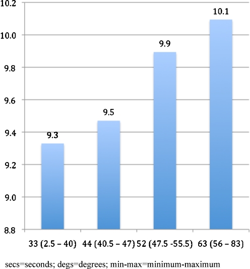
Mean mobility time increased from 9.3 s in the lowest to 10.1 s in the highest quartile of kyphosis. In a multivariate-adjusted model, mobility time increased 0.11 s (p = 0.02) for each standard deviation (11.9°) increase in kyphosis. Longer performance times were significantly associated with increasing age, decreasing grip strength, vertebral fractures, body mass index ≥25, and total hip BMD in the osteoporotic range.
Kyphosis angle is independently associated with decreased mobility on the Timed Up and Go, which is in turn correlated with increased fall risk. Hyperkyphosis may be a useful clinical marker signaling the need for evaluation of vertebral fracture and falling risk.
本研究旨在确定脊柱后凸是否独立于骨质疏松症与移动能力相关,以及量化其他导致移动能力受损的因素的影响。
我们使用了 1993 年基线访视时年龄在 55-80 岁之间的 3108 名社区居住的女性参与者的数据。所有参与者都进行了脊柱后凸、计时起立行走测试的移动时间、身高、体重、全髋骨密度(BMD)、握力和椎体骨折的测量。人口统计学特征包括年龄和吸烟状况。我们按脊柱后凸的四分位数计算了平均计时起立行走测试时间。使用多元线性回归,我们估计了脊柱后凸与移动时间的独立关联,并量化了其他协变量对移动能力的影响。
移动时间从脊柱后凸最低四分位数的 9.3 秒增加到最高四分位数的 10.1 秒。在多元调整模型中,脊柱后凸每增加一个标准差(11.9°),移动时间增加 0.11 秒(p=0.02)。较长的运动时间与年龄增加、握力下降、椎体骨折、体重指数≥25 和全髋 BMD 在骨质疏松范围内显著相关。
脊柱后凸角度与计时起立行走测试的移动能力下降独立相关,而移动能力下降又与跌倒风险增加相关。脊柱后凸可能是一个有用的临床标志物,表明需要评估椎体骨折和跌倒风险。