Center for Imaging of Neurodegenerative Diseases, Veterans Affairs Medical Center, Department of Radiology and Biomedical Imaging, University of California, San Francisco, 4150 Clement Street, 114M, San Francisco, CA 94121, USA.
Brain Imaging Behav. 2010 Mar;4(1):46-54. doi: 10.1007/s11682-009-9084-1.
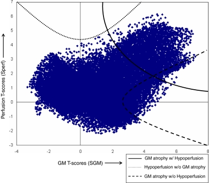
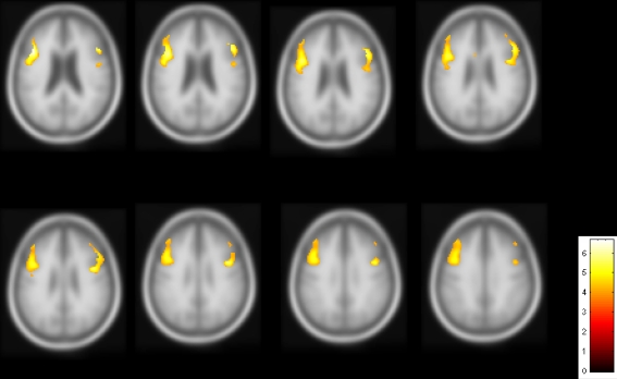
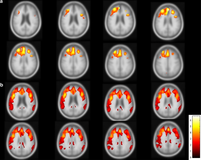
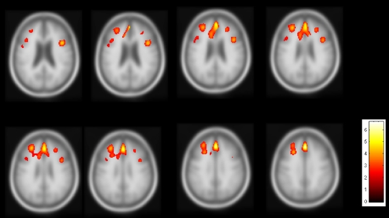
The aim of this study was to determine if a dissociation between reduced cerebral perfusion and gray matter (GM) atrophy exists in frontotemporal dementia (FTD). The study included 28 patients with FTD and 29 cognitive normal (CN) subjects. All subjects had MRI at 1.5 T, including T1-weighted structural and arterial spin labeling (ASL) perfusion imaging. Non-parametric concordance/discordance tests revealed that GM atrophy without hypoperfusion occurs in the premotor cortex in FTD whereas concordant GM atrophy and hypoperfusion changes are found in the right prefrontal cortex and bilateral medial frontal lobe. The results suggest that damage of brain function in FTD, assessed by ASL perfusion, can vary regionally despite widespread atrophy. Detection of discordance between brain perfusion and structure in FTD might aid diagnosis and staging of the disease.
本研究旨在确定额颞叶痴呆(FTD)是否存在脑灌注减少与灰质(GM)萎缩分离的现象。该研究纳入了 28 例 FTD 患者和 29 例认知正常(CN)受试者。所有受试者均在 1.5T 磁共振仪上进行 MRI 检查,包括 T1 加权结构像和动脉自旋标记(ASL)灌注成像。非参数一致性/不一致性检验显示,FTD 患者的运动前皮质存在无灌注性 GM 萎缩,而在右侧前额叶皮质和双侧额内侧回则存在 GM 萎缩和灌注改变一致的现象。这些结果表明,FTD 患者的脑功能损伤可以通过 ASL 灌注来评估,尽管存在广泛的萎缩,但损伤的区域可能会有所不同。在 FTD 中检测脑灌注与结构之间的不一致可能有助于该疾病的诊断和分期。