Christov K, Chew K L, Ljung B M, Waldman F M, Duarte L A, Goodson W H, Smith H S, Mayall B H
Department of Laboratory Medicine, University of California, San Francisco 94143-0808.
Am J Pathol. 1991 Jun;138(6):1371-7.
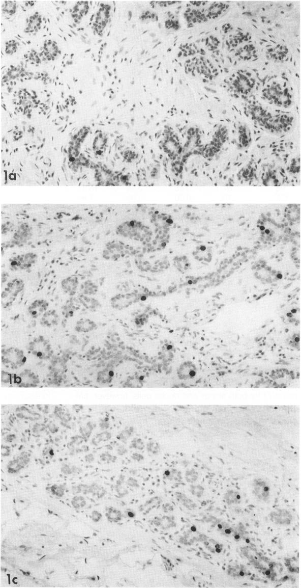
The proliferative activity of normal acinar and ductal breast epithelial cells was studied by in vivo labeling with 5-bromodeoxyuridine (BrdUrd) in 26 cases with concurrent breast carcinoma. The BrdUrd-labeled cells were recognized in histologic sections of paraffin-embedded tissue, using an anti-BrdUrd antibody and an immunoperoxidase reaction. The percentage of BrdUrd-labeled cells showed great variability for both acinar (0% to 2.66%; mean, 0.70%; standard deviation [SD], 0.80%) and ductal cells (0% to 1.99%; mean, 0.51%; SD, 0.57%). The fraction of proliferating epithelial cells declined with the age of the patients and was significantly higher in premenopausal women (1.16% +/- 0.85% for acinar and 0.94% +/- 0.60% for ductal cells) as compared with the postmenopausal women (0.27% +/- 0.46% for acinar and 0.17% +/- 0.22% for ductal cells), P less than 0.01 for acinar and P less than 0.001 for ductal cells, respectively. In some patients, great variability in distribution of proliferating acinar and ductal cells among different lobules and ducts was observed. No difference was found in the number of proliferating acinar and ductal cells situated near or far from their corresponding tumors. No correlation was seen between cell proliferation of normal acinar or ductal cells and cell proliferation of the respective tumors.
通过对26例同时患有乳腺癌的患者进行5-溴脱氧尿苷(BrdUrd)体内标记,研究了正常乳腺腺泡和导管上皮细胞的增殖活性。使用抗BrdUrd抗体和免疫过氧化物酶反应,在石蜡包埋组织的组织学切片中识别出BrdUrd标记的细胞。BrdUrd标记细胞的百分比在腺泡细胞(0%至2.66%;平均值为0.70%;标准差[SD]为0.80%)和导管细胞(0%至1.99%;平均值为0.51%;SD为0.57%)中均表现出很大的变异性。增殖上皮细胞的比例随患者年龄的增加而下降,绝经前女性(腺泡细胞为1.16%±0.85%,导管细胞为0.94%±0.60%)的比例明显高于绝经后女性(腺泡细胞为0.27%±0.46%,导管细胞为0.17%±0.22%),腺泡细胞P<0.01,导管细胞P<0.001。在一些患者中,观察到不同小叶和导管中增殖腺泡和导管细胞分布的很大变异性。在距相应肿瘤近或远的位置,增殖腺泡和导管细胞的数量没有差异。正常腺泡或导管细胞的细胞增殖与相应肿瘤的细胞增殖之间没有相关性。