• 文献检索
  • 文档翻译
  • 深度研究
  • 学术资讯
  • Suppr Zotero 插件Zotero 插件
  • 邀请有礼
  • 套餐&价格
  • 历史记录
应用&插件
Suppr Zotero 插件Zotero 插件浏览器插件Mac 客户端Windows 客户端微信小程序
定价
高级版会员购买积分包购买API积分包
服务
文献检索文档翻译深度研究API 文档MCP 服务
关于我们
关于 Suppr公司介绍联系我们用户协议隐私条款
关注我们

Suppr 超能文献

核心技术专利:CN118964589B侵权必究
粤ICP备2023148730 号-1Suppr @ 2026

文献检索

告别复杂PubMed语法,用中文像聊天一样搜索,搜遍4000万医学文献。AI智能推荐,让科研检索更轻松。

立即免费搜索

文件翻译

保留排版,准确专业,支持PDF/Word/PPT等文件格式,支持 12+语言互译。

免费翻译文档

深度研究

AI帮你快速写综述,25分钟生成高质量综述,智能提取关键信息,辅助科研写作。

立即免费体验

量子点标记的舌鳞癌细胞中窖蛋白-1 的表达

Expression of caveolin-1 in tongue squamous cell carcinoma by quantum dots.

机构信息

Department of Pathology, School of Basic Medical Science, Wuhan University, Wuhan, China.

出版信息

Eur J Histochem. 2010 Apr 14;54(2):e20. doi: 10.4081/ejh.2010.e20.

DOI:10.4081/ejh.2010.e20
PMID:20558341
原文链接:https://pmc.ncbi.nlm.nih.gov/articles/PMC3167300/
Abstract

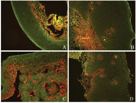
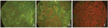
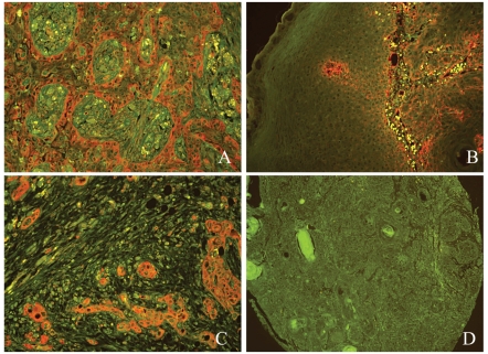
Quantum dots (QDs) are a new class of fluorescent probes to detect biomarker expression. The role of caveolin-1 (Cav-1) in tongue squamous cell carcinoma (TSCC) is still unknown. This study aimed to investigate the expression profile of Cav-1 in carcinogenesis and development of TSCC by QDs immunofluorescence histochemistry (QDs-IHC) and discuss the relationship between the Cav-1 expression and the clinicopathological outcomes. QDs-IHC was used to detect Cav-1 expression in tissue microarrays including normal tongue mucosa (NTM; n=10), hyperplastic tongue mucosa (HTM; n=10), tongue pre-cancer lesions (TPL; n=15) and primary tongue squamous cell carcinoma (PTSCC; n=61). Correlations between the Cav-1 expression and clinicopathologic variables were evaluated statistically. Cells positive for Cav-1 were clearly detected and bright images were obtained in a fine, granular pattern at the cell membrane and cytoplasm using QDs-IHC. The rate of Cav-1 immunoreactivity increased progressively from NTM (0%), HTM (0%), TPL (36%) to PTSCC (74%). When compared with each other, there was statistical significance among PTSCC, TPL and NTM as well as among PTSCC, TPL and HTM. Moreover, Cav-1 expression level in PTSCC was correlated positively with clinical stage and histologic grade. QDs-IHC could accurately detect protein location in tongue mucosa. An increased expression of Cav-1 in the stepwise carcinogenesis from NTM, HTM, TPL to PTSCC suggested that Cav-1 might be an oncogene in the development of tongue squamous cell carcinoma.

摘要

量子点 (QDs) 是一种新型荧光探针,用于检测生物标志物的表达。窖蛋白-1 (Cav-1) 在舌鳞状细胞癌 (TSCC) 中的作用尚不清楚。本研究旨在通过 QDs 免疫荧光组织化学 (QDs-IHC) 检测 Cav-1 在 TSCC 发生和发展过程中的表达谱,并探讨 Cav-1 表达与临床病理结局的关系。QDs-IHC 用于检测组织微阵列中 Cav-1 的表达,包括正常舌黏膜 (NTM; n=10)、增生性舌黏膜 (HTM; n=10)、舌前病变 (TPL; n=15) 和原发性舌鳞状细胞癌 (PTSCC; n=61)。统计评估 Cav-1 表达与临床病理变量之间的相关性。使用 QDs-IHC,细胞膜和细胞质中 Cav-1 阳性细胞清晰可见,呈细颗粒状明亮图像。Cav-1 免疫反应率从 NTM(0%)、HTM(0%)、TPL(36%)逐渐增加到 PTSCC(74%)。与彼此相比,PTSCC、TPL 和 NTM 之间以及 PTSCC、TPL 和 HTM 之间均有统计学意义。此外,PTSCC 中 Cav-1 的表达水平与临床分期和组织学分级呈正相关。QDs-IHC 可以准确检测舌黏膜中蛋白质的位置。Cav-1 在从 NTM、HTM、TPL 到 PTSCC 的逐步癌变过程中的表达增加表明,Cav-1 可能是舌鳞状细胞癌发生的癌基因。

https://cdn.ncbi.nlm.nih.gov/pmc/blobs/fcee/3167300/b56d5d71f913/ejh-2010-2-e20-g003.jpg
https://cdn.ncbi.nlm.nih.gov/pmc/blobs/fcee/3167300/509f2520d97c/ejh-2010-2-e20-g001.jpg
https://cdn.ncbi.nlm.nih.gov/pmc/blobs/fcee/3167300/340c21cd8907/ejh-2010-2-e20-g002.jpg
https://cdn.ncbi.nlm.nih.gov/pmc/blobs/fcee/3167300/b56d5d71f913/ejh-2010-2-e20-g003.jpg
https://cdn.ncbi.nlm.nih.gov/pmc/blobs/fcee/3167300/509f2520d97c/ejh-2010-2-e20-g001.jpg
https://cdn.ncbi.nlm.nih.gov/pmc/blobs/fcee/3167300/340c21cd8907/ejh-2010-2-e20-g002.jpg
https://cdn.ncbi.nlm.nih.gov/pmc/blobs/fcee/3167300/b56d5d71f913/ejh-2010-2-e20-g003.jpg

相似文献

1
Expression of caveolin-1 in tongue squamous cell carcinoma by quantum dots.量子点标记的舌鳞癌细胞中窖蛋白-1 的表达
Eur J Histochem. 2010 Apr 14;54(2):e20. doi: 10.4081/ejh.2010.e20.
2
[Significance of caveolin-1 protein detected by quantum dots technique in human lung cancer invasiveness and metastasis.].
Zhonghua Jie He He Hu Xi Za Zhi. 2009 Nov;32(11):821-4.
3
NIT2 overexpression predicts poor prognosis in tongue squamous cell carcinoma patients.NIT2 过表达预示着舌鳞癌患者预后不良。
Mol Biol Rep. 2020 Mar;47(3):1553-1561. doi: 10.1007/s11033-019-05197-5. Epub 2020 Jan 10.
4
Quantum dots-based immunofluorescent imaging of stromal fibroblasts Caveolin-1 and light chain 3B expression and identification of their clinical significance in human gastric cancer.基于量子点的基质成纤维细胞小窝蛋白-1和轻链3B表达的免疫荧光成像及其在人胃癌中的临床意义鉴定
Int J Mol Sci. 2012 Oct 24;13(11):13764-80. doi: 10.3390/ijms131113764.
5
Caveolin-1 accumulation in the tongue cancer tumor microenvironment is significantly associated with poor prognosis: an in-vivo and in-vitro study.小窝蛋白-1在舌癌肿瘤微环境中的积聚与预后不良显著相关:一项体内和体外研究。
BMC Cancer. 2015 Jan 30;15:25. doi: 10.1186/s12885-015-1030-6.
6
Differences in Clinicopathological Characteristics and Prognosis Between Primary and Postirradiation Tongue Squamous Cell Carcinoma.原发性与放疗后舌鳞状细胞癌的临床病理特征及预后差异
J Oral Maxillofac Surg. 2017 Oct;75(10):2235-2241. doi: 10.1016/j.joms.2017.02.020. Epub 2017 Mar 6.
7
Expression of Cav-1 in tumour cells, rather than in stromal tissue, may promote cervical squamous cell carcinoma proliferation, and correlates with high-risk HPV infection.Cav-1 在肿瘤细胞中的表达,而不是在基质组织中的表达,可能促进宫颈鳞状细胞癌的增殖,并与高危 HPV 感染相关。
Oncol Rep. 2012 Jun;27(6):1733-40. doi: 10.3892/or.2012.1703. Epub 2012 Feb 28.
8
Comparison of quantum dots immunofluorescence histochemistry and conventional immunohistochemistry for the detection of caveolin-1 and PCNA in the lung cancer tissue microarray.量子点免疫荧光组织化学法与常规免疫组织化学法检测肺癌组织芯片中小窝蛋白-1和 PCNA 的比较。
J Mol Histol. 2009 Aug;40(4):261-8. doi: 10.1007/s10735-009-9237-y. Epub 2009 Nov 12.
9
Global DNA hypomethylation is associated with the development and poor prognosis of tongue squamous cell carcinoma.全基因组DNA低甲基化与舌鳞状细胞癌的发生发展及不良预后相关。
J Oral Pathol Med. 2016 Jul;45(6):409-17. doi: 10.1111/jop.12381. Epub 2015 Nov 2.
10
Loss of Caveolin-1 Expression in Tumor Cells is Associated with Increased Aggressiveness and Cell Invasion in Oral Squamous Cell Carcinoma.肿瘤细胞中窖蛋白-1 表达缺失与口腔鳞状细胞癌侵袭性和细胞迁移增加相关。
Head Neck Pathol. 2023 Sep;17(3):618-630. doi: 10.1007/s12105-023-01562-w. Epub 2023 May 26.

引用本文的文献

1
Multi-omics exploration of CAV1 tumor epithelial subcelltype in oral squamous cell carcinoma and its impact on the immune microenvironment.口腔鳞状细胞癌中CAV1肿瘤上皮亚细胞类型的多组学探索及其对免疫微环境的影响
Discov Oncol. 2025 Jun 5;16(1):1006. doi: 10.1007/s12672-025-02843-2.
2
Increased Plasmatic Levels of Exosomes Are Significantly Related to Relapse Rate in Patients with Oral Squamous Cell Carcinoma: A Cohort Study.血浆中外泌体水平升高与口腔鳞状细胞癌患者的复发率显著相关:一项队列研究。
Cancers (Basel). 2023 Dec 2;15(23):5693. doi: 10.3390/cancers15235693.
3
Cholesterol Is a Regulator of CAV1 Localization and Cell Migration in Oral Squamous Cell Carcinoma.

本文引用的文献

1
Comparison of quantum dots immunofluorescence histochemistry and conventional immunohistochemistry for the detection of caveolin-1 and PCNA in the lung cancer tissue microarray.量子点免疫荧光组织化学法与常规免疫组织化学法检测肺癌组织芯片中小窝蛋白-1和 PCNA 的比较。
J Mol Histol. 2009 Aug;40(4):261-8. doi: 10.1007/s10735-009-9237-y. Epub 2009 Nov 12.
2
Caveolin-1 overexpression correlates with tumour progression markers in pancreatic ductal adenocarcinoma.小窝蛋白-1过表达与胰腺导管腺癌中的肿瘤进展标志物相关。
J Mol Histol. 2009 Feb;40(1):23-9. doi: 10.1007/s10735-008-9209-7. Epub 2009 Jan 22.
3
Restoration of caveolin-1 expression suppresses growth and metastasis of head and neck squamous cell carcinoma.
胆固醇是口腔鳞状细胞癌中 Cav1 定位和细胞迁移的调节剂。
Int J Mol Sci. 2023 Mar 23;24(7):6035. doi: 10.3390/ijms24076035.
4
Identification of Potential Biomarkers Using Integrative Approach: A Case Study of ESCC.使用综合方法鉴定潜在生物标志物:以食管癌为例
SN Comput Sci. 2023;4(2):114. doi: 10.1007/s42979-022-01492-4. Epub 2022 Dec 21.
5
Involvement of caveolin-1 in skin diseases.窖蛋白-1 在皮肤疾病中的作用。
Front Immunol. 2022 Nov 30;13:1035451. doi: 10.3389/fimmu.2022.1035451. eCollection 2022.
6
Airway basal cells show regionally distinct potential to undergo metaplastic differentiation.气道基底细胞表现出区域特有的多能性分化潜能。
Elife. 2022 Sep 30;11:e80083. doi: 10.7554/eLife.80083.
7
Quantitative proteomics identifies biomarkers to distinguish pulmonary from head and neck squamous cell carcinomas by immunohistochemistry.定量蛋白质组学通过免疫组织化学鉴定用于区分肺和头颈部鳞状细胞癌的生物标志物。
J Pathol Clin Res. 2022 Jan;8(1):33-47. doi: 10.1002/cjp2.244. Epub 2021 Oct 14.
8
Nanoparticles for Oral Cancer Diagnosis and Therapy.用于口腔癌诊断与治疗的纳米颗粒
Bioinorg Chem Appl. 2021 Apr 23;2021:9977131. doi: 10.1155/2021/9977131. eCollection 2021.
9
Differences in the expression of caveolin-1 isoforms in cancer-associated and normal fibroblasts of patients with oral squamous cell carcinoma.口腔鳞状细胞癌患者癌相关成纤维细胞与正常成纤维细胞中窖蛋白-1 异构体表达的差异。
Clin Oral Investig. 2021 Oct;25(10):5823-5831. doi: 10.1007/s00784-021-03887-8. Epub 2021 Mar 27.
10
Identification of four key prognostic genes and three potential drugs in human papillomavirus negative head and neck squamous cell carcinoma.人乳头瘤病毒阴性头颈部鳞状细胞癌中四个关键预后基因和三种潜在药物的鉴定
Cancer Cell Int. 2021 Mar 12;21(1):167. doi: 10.1186/s12935-021-01863-6.
小窝蛋白-1表达的恢复可抑制头颈部鳞状细胞癌的生长和转移。
Br J Cancer. 2008 Nov 18;99(10):1684-94. doi: 10.1038/sj.bjc.6604735. Epub 2008 Oct 28.
4
Caveolin-1 expression in human breast lobular cancer progression.小窝蛋白-1在人乳腺小叶癌进展中的表达
Mod Pathol. 2009 Jan;22(1):71-8. doi: 10.1038/modpathol.2008.154. Epub 2008 Oct 3.
5
In vivo biodistribution and highly efficient tumour targeting of carbon nanotubes in mice.碳纳米管在小鼠体内的生物分布及高效肿瘤靶向性
Nat Nanotechnol. 2007 Jan;2(1):47-52. doi: 10.1038/nnano.2006.170. Epub 2006 Dec 17.
6
The overexpression of caveolin-1 and caveolin-2 correlates with a poor prognosis and tumor progression in esophageal squamous cell carcinoma.小窝蛋白-1和小窝蛋白-2的过表达与食管鳞状细胞癌的预后不良和肿瘤进展相关。
Oncol Rep. 2007 Sep;18(3):601-9.
7
Bioconjugated quantum dots for multiplexed and quantitative immunohistochemistry.用于多重定量免疫组织化学的生物共轭量子点
Nat Protoc. 2007;2(5):1152-65. doi: 10.1038/nprot.2007.107.
8
Caveolin-1 overexpression is associated with aggressive prostate cancer recurrence.小窝蛋白-1过表达与侵袭性前列腺癌复发相关。
Prostate. 2007 May 1;67(6):614-22. doi: 10.1002/pros.20557.
9
Semiautomated multiplexed quantum dot-based in situ hybridization and spectral deconvolution.基于量子点的半自动多重原位杂交及光谱解卷积
J Mol Diagn. 2007 Feb;9(1):20-9. doi: 10.2353/jmoldx.2007.060119.
10
Caveolin 1 is overexpressed and amplified in a subset of basal-like and metaplastic breast carcinomas: a morphologic, ultrastructural, immunohistochemical, and in situ hybridization analysis.小窝蛋白1在一部分基底样和化生性乳腺癌中过表达且扩增:形态学、超微结构、免疫组织化学及原位杂交分析
Clin Cancer Res. 2007 Jan 1;13(1):90-101. doi: 10.1158/1078-0432.CCR-06-1371.