Department of Radiology and Diagnostic Imaging, Regina Elena Institute, E, Chianesi 53, Rome, Italy.
J Exp Clin Cancer Res. 2010 Jun 17;29(1):73. doi: 10.1186/1756-9966-29-73.
To retrospectively compare the diagnostic accuracy of magnetic resonance imaging (MRI) and multidetector-row computed tomography (MDCT) in the assessment of the mandibular invasion by squamous cell carcinoma (SCC) having histopathological exams as standard of reference.
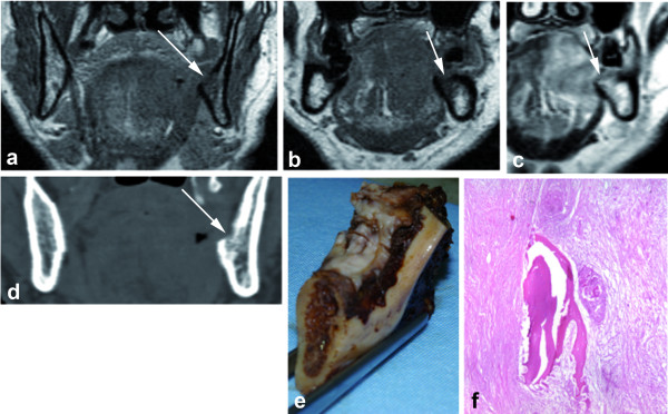
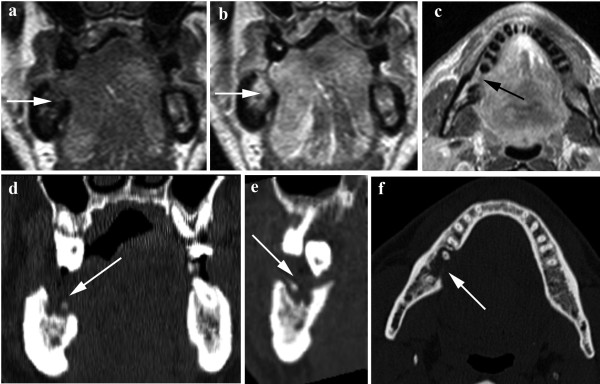
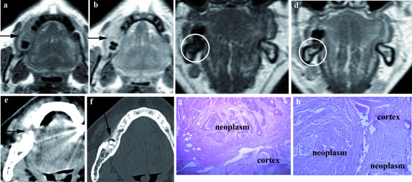
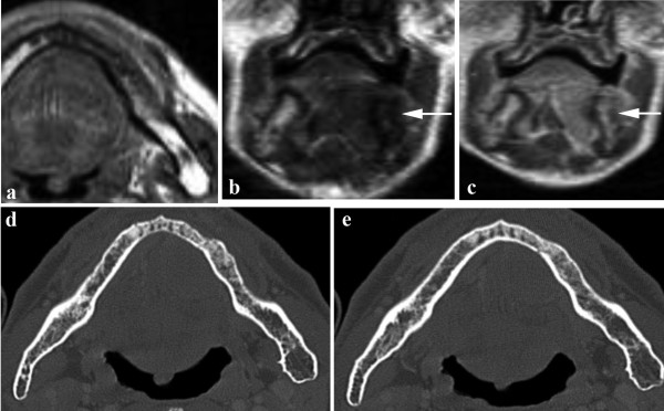
Institutional review board approval with a waiver of informed patient consent was obtained. Of the 147 patients selected from our database who underwent surgical excision of a tumour arising into the oral cavity, thirty-six patients (26 men, 10 women; mean age, 56 years; range, 30-75 years) with hystologically proven SCC who performed both a preoperative MRI and MDCT, composed our final study population.Images were qualitatively analyzed in consensus by two expert radiologist in head and neck imaging. Sensitivity, specificity, accuracy, positive predictive value (PPV) and negative predictive value (NPV) were assessed for both MRI and MDCT.Differences in sensitivity, specificity, positive and negative predictive values were calculated at a statistical significance of p < .05.
The sensitivity, the specificity and the accuracy of MRI and MDCT in the detection of the mandibular involvement were respectively 93%, 82%, 86% and 79%, 82%, 81%, while the positive predictive value (PPV) and negative predictive value (NPV) were respectively 76%, 95% and 73%, 86%. There wasn't any statistically significant difference in overall diagnostic accuracy between MRI and MDCT in the evaluation of mandibular tumour invasion (p > .05).
MRI showed to have a higher sensitivity compare to MDCT in the assessment of mandibular involvement from SCC arising in the oral cavity although none statistically significant differences were noted.
回顾性比较磁共振成像(MRI)和多层螺旋 CT(MDCT)在评估鳞癌(SCC)侵犯下颌骨方面的诊断准确性,以组织病理学检查为参考标准。
获得机构审查委员会批准并豁免患者知情同意。从我们的数据库中选择了 147 名接受口腔肿瘤切除术的患者,其中 36 名(26 名男性,10 名女性;平均年龄 56 岁;范围 30-75 岁)经组织学证实为 SCC,术前均行 MRI 和 MDCT 检查,构成了我们的最终研究人群。图像由两名头颈成像专家进行定性分析。评估了 MRI 和 MDCT 对下颌骨侵犯的敏感性、特异性、准确性、阳性预测值(PPV)和阴性预测值(NPV)。在统计学意义上(p<.05)计算了敏感性、特异性、阳性和阴性预测值的差异。
MRI 和 MDCT 检测下颌骨受累的敏感性、特异性和准确性分别为 93%、82%、86%和 79%、82%、81%,阳性预测值(PPV)和阴性预测值(NPV)分别为 76%、95%和 73%、86%。在评估口腔 SCC 下颌骨侵犯方面,MRI 和 MDCT 在整体诊断准确性方面没有统计学上的显著差异(p>.05)。
与 MDCT 相比,MRI 在评估 SCC 侵犯下颌骨方面具有更高的敏感性,但无统计学差异。