Working Group on Cardiovascular Magnetic Resonance, Medical University Berlin, Charité Campus Buch, HELIOS Klinikum Berlin-Buch, Department of Cardiology and Nephrology, Schwanebecker Chaussee 50, 13125 Berlin, Germany.
J Cardiovasc Magn Reson. 2010 Jun 23;12(1):36. doi: 10.1186/1532-429X-12-36.
The orifice area of mitral bioprostheses provides important information regarding their hemodynamic performance. It is usually calculated by transthoracic echocardiography (TTE), however, accurate and reproducible determination may be challenging. Cardiovascular magnetic resonance (CMR) has been proven as an accurate alternative for assessing aortic bioprostheses. However, whether CMR can be similarly applied for bioprostheses in the mitral position, particularly in the presence of frequently coincident arrhythmias, is unclear. The aim of the study is to test the feasibility of CMR to evaluate the orifice area of mitral bioprostheses.
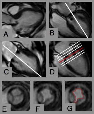
CMR planimetry was performed in 18 consecutive patients with mitral bioprostheses (n = 13 Hancock(R), n = 4 Labcore(R), n = 1 Perimount(R); mean time since implantation 4.5 +/- 3.9 years) in an imaging plane perpendicular to the transprosthetic flow using steady-state free-precession cine imaging under breath-hold conditions on a 1.5T MR system. CMR results were compared with pressure half-time derived orifice areas obtained by TTE.
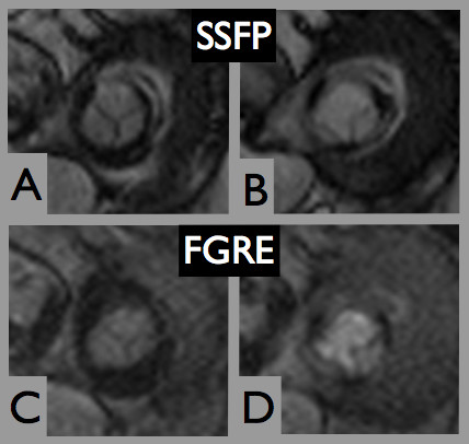
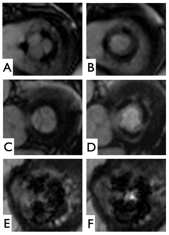
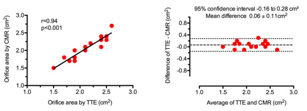
Six subjects were in sinus rhythm, 11 in atrial fibrillation, and 1 exhibited frequent ventricular extrasystoles. CMR image quality was rated as good in 10, moderate in 6, and significantly impaired in 2 subjects. In one prosthetic type (Perimount(R)), strong stent artifacts occurred. Orifice areas by CMR (mean 2.1 +/- 0.3 cm2) and TTE (mean 2.1 +/- 0.3 cm2) correlated significantly (r = 0.94; p < 0.001). Bland-Altman analysis showed a 95% confidence interval from -0.16 to 0.28 cm2 (mean difference 0.06 +/- 0.11 cm2; range -0.1 to 0.3 cm2). Intra- and inter-observer variabilities of CMR planimetry were 4.5 +/- 2.9% and 7.9 +/- 5.2%.
The assessment of mitral bioprostheses using CMR is feasible even in those with arrhythmias, providing orifice areas with close agreement to echocardiography and low observer dependency. Larger samples with a greater variety of prosthetic types and more cases of prosthetic dysfunction are required to confirm these preliminary results.
二尖瓣生物瓣的瓣口面积提供了有关其血流动力学性能的重要信息。通常通过经胸超声心动图(TTE)进行计算,但是,准确且可重复的确定可能具有挑战性。心血管磁共振(CMR)已被证明是评估主动脉生物瓣的准确替代方法。然而,CMR 是否可以类似地应用于二尖瓣位置的生物瓣,特别是在经常同时存在心律失常的情况下,尚不清楚。该研究的目的是测试 CMR 评估二尖瓣生物瓣瓣口面积的可行性。
在 1.5T MR 系统上使用稳态自由进动电影成像在与跨瓣血流垂直的成像平面上对 18 例连续接受二尖瓣生物瓣(n = 13 例 Hancock(R),n = 4 例 Labcore(R),n = 1 例 Perimount(R))的患者进行 CMR 平面测量,在屏气条件下进行。将 CMR 结果与 TTE 获得的压力半衰期衍生瓣口面积进行比较。
6 例患者处于窦性心律,11 例患者处于心房颤动,1 例患者频发室性早搏。10 例患者的 CMR 图像质量评为良好,6 例患者评为中度,2 例患者评为明显受损。在一种假体类型(Perimount(R))中,出现了强烈的支架伪影。CMR(平均 2.1 +/- 0.3 cm2)和 TTE(平均 2.1 +/- 0.3 cm2)的瓣口面积显著相关(r = 0.94;p < 0.001)。Bland-Altman 分析显示 95%置信区间为-0.16 至 0.28 cm2(平均差值 0.06 +/- 0.11 cm2;范围-0.1 至 0.3 cm2)。CMR 平面测量的内和观察者间变异性分别为 4.5 +/- 2.9%和 7.9 +/- 5.2%。
即使在存在心律失常的情况下,使用 CMR 评估二尖瓣生物瓣也是可行的,提供的瓣口面积与超声心动图非常吻合,且观察者依赖性低。需要更大的样本量、更多种类的假体和更多假体功能障碍的病例来证实这些初步结果。