Department of Anesthesia and Critical Care, Beth Israel Deaconess Medical Center, Boston, MA.
Ann Neurol. 2010 Jul;68(1):81-91. doi: 10.1002/ana.21994.
Focal somatic pain can evolve into widespread hypersensitivity to nonpainful and painful skin stimuli (allodynia and hyperalgesia, respectively). We hypothesized that transformation of headache into whole-body allodynia/hyperalgesia during a migraine attack is mediated by sensitization of thalamic neurons that process converging sensory impulses from the cranial meninges and extracephalic skin.
Extracephalic allodynia was assessed using single unit recording of thalamic trigeminovascular neurons in rats and contrast analysis of blood oxygenation level-dependent (BOLD) signals registered in functional magnetic resonance imaging (fMRI) scans of patients exhibiting extracephalic allodynia.
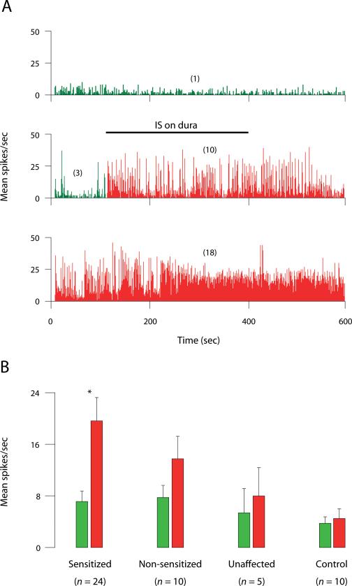
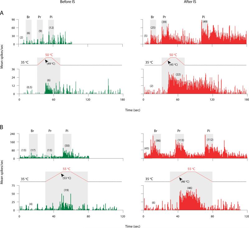
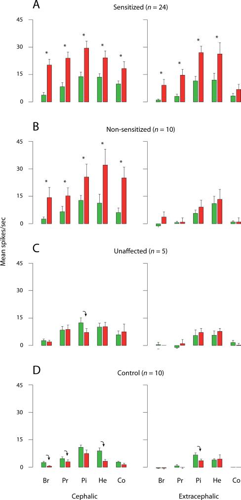
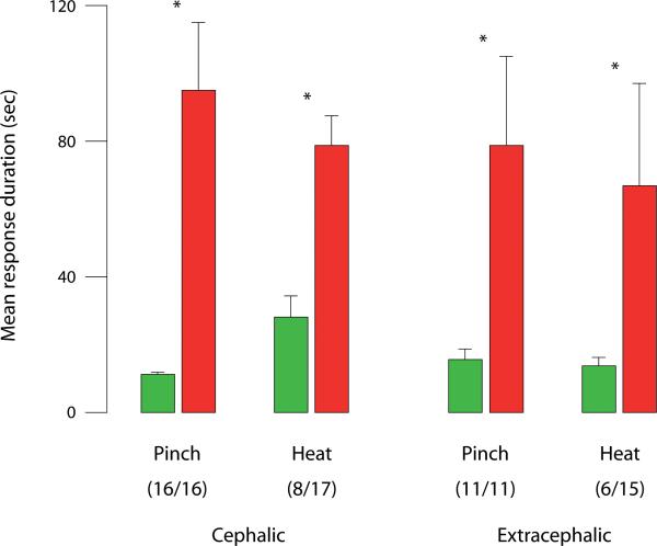
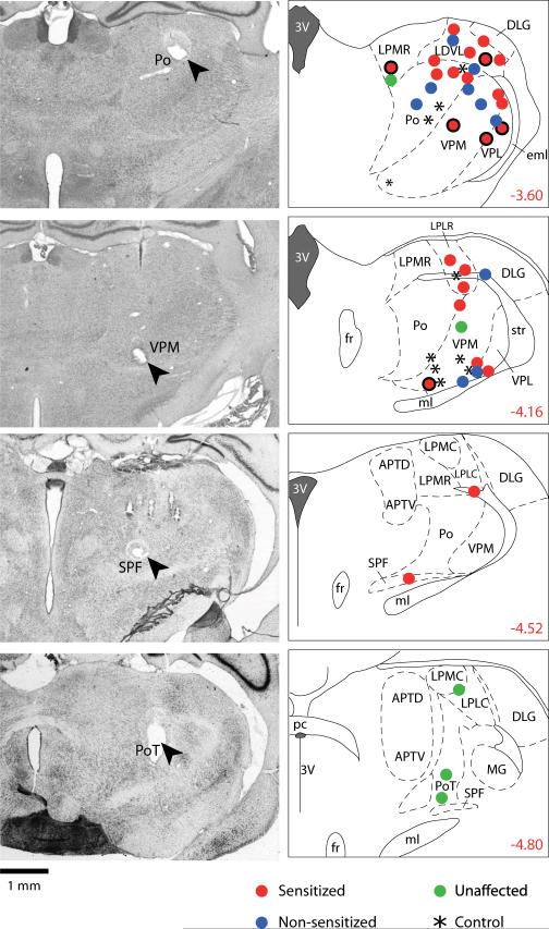
Sensory neurons in the rat posterior thalamus that were activated and sensitized by chemical stimulation of the cranial dura exhibited long-lasting hyperexcitability to innocuous (brush, pressure) and noxious (pinch, heat) stimulation of the paws. Innocuous, extracephalic skin stimuli that did not produce neuronal firing at baseline (eg, brush) became as effective as noxious stimuli (eg, pinch) in eliciting large bouts of neuronal firing after sensitization was established. In migraine patients, fMRI assessment of BOLD signals showed that brush and heat stimulation at the skin of the dorsum of the hand produced larger BOLD responses in the posterior thalamus of subjects undergoing a migraine attack with extracephalic allodynia than the corresponding responses registered when the same patients were free of migraine and allodynia.
We propose that the spreading of multimodal allodynia and hyperalgesia beyond the locus of migraine headache is mediated by sensitized thalamic neurons that process nociceptive information from the cranial meninges together with sensory information from the skin of the scalp, face, body, and limbs.
躯体局部疼痛可演变为对非疼痛和疼痛皮肤刺激的广泛超敏反应(分别为痛觉过敏和痛觉过敏)。我们假设偏头痛发作期间头痛向全身痛觉过敏/痛觉过敏的转变是由处理来自颅脑膜和颅外皮肤的汇聚感觉冲动的丘脑神经元敏化介导的。
使用大鼠三叉血管神经元的单个单位记录评估颅外痛觉过敏,并对比分析表现出颅外痛觉过敏的患者的功能磁共振成像(fMRI)扫描中记录的血氧水平依赖(BOLD)信号。
化学刺激颅硬膜激活和敏化大鼠后丘脑的感觉神经元对足部的无害(刷,压)和有害(捏,热)刺激表现出持久的超兴奋性。在基线时不会产生神经元放电的无害的颅外皮肤刺激物(例如,刷)在敏化建立后变得与有害刺激物(例如,捏)一样有效地引起大量神经元放电。在偏头痛患者中,BOLD 信号的 fMRI 评估显示,在手背部皮肤的刷和热刺激在经历颅外痛觉过敏的偏头痛发作的受试者的后丘脑产生比在相同患者没有偏头痛和痛觉过敏时记录的相应反应更大的 BOLD 反应。
我们提出,多模态痛觉过敏和痛觉过敏的扩散超出偏头痛头痛的部位是由处理来自颅脑膜的伤害性信息以及来自头皮,面部,身体和四肢的感觉信息的敏化丘脑神经元介导的。