Department of Pharmacology, University of Arizona College of Medicine, 1501 N Campbell Ave, PO Box 245050, Tucson, AZ 85724, USA.
Mol Pain. 2012 Jan 24;8:6. doi: 10.1186/1744-8069-8-6.
Migraine headache is one of the most common neurological disorders, but the pathophysiology contributing to migraine is poorly understood. Intracranial interleukin-6 (IL-6) levels have been shown to be elevated during migraine attacks, suggesting that this cytokine may facilitate pain signaling from the meninges and contribute to the development of headache.
Cutaneous allodynia was measured in rats following stimulation of the dura with IL-6 alone or in combination with the MEK inhibitor, U0126. The number of action potentials and latency to the first action potential peak in response to a ramp current stimulus as well as current threshold were measured in retrogradely-labeled dural afferents using patch-clamp electrophysiology. These recordings were performed in the presence of IL-6 alone or in combination with U0126. Association between ERK1 and Nav1.7 following IL-6 treatment was also measured by co-immunoprecipitation.
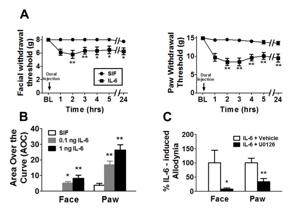
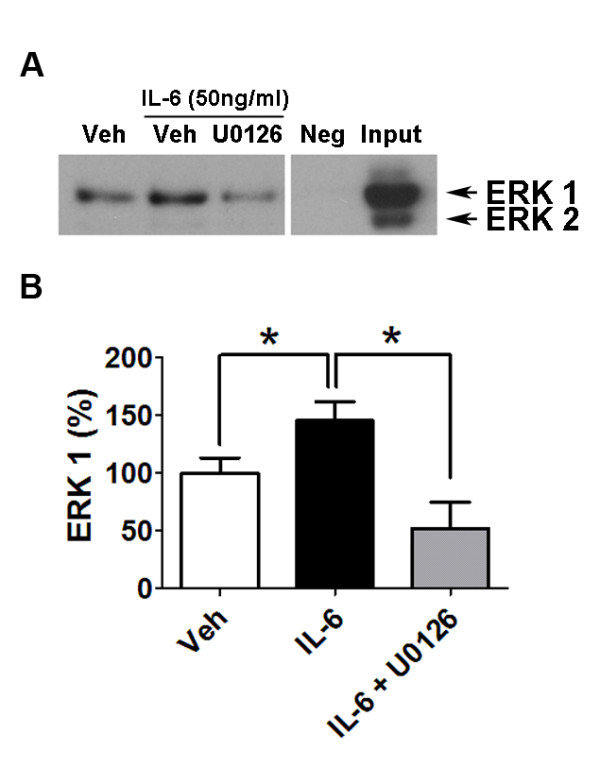
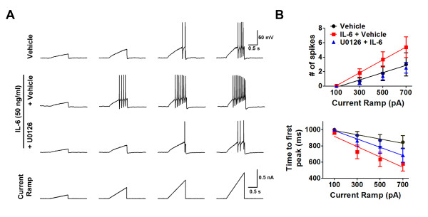
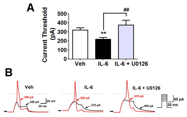
Here we report that in awake animals, direct application of IL-6 to the dura produced dose-dependent facial and hindpaw allodynia. The MEK inhibitor U0126 blocked IL-6-induced allodynia indicating that IL-6 produced this behavioral effect through the MAP kinase pathway. In trigeminal neurons retrogradely labeled from the dura, IL-6 application decreased the current threshold for action potential firing. In response to a ramp current stimulus, cells treated with IL-6 showed an increase in the numbers of action potentials and a decrease in latency to the first spike, an effect consistent with phosphorylation of the sodium channel Nav1.7. Pretreatment with U0126 reversed hyperexcitability following IL-6 treatment. Moreover, co-immunoprecipitation experiments demonstrated an increased association between ERK1 and Nav1.7 following IL-6 treatment.
Our results indicate that IL-6 enhances the excitability of dural afferents likely via ERK-mediated modulation of Nav1.7 and these responses contribute to migraine-related pain behavior in vivo. These data provide a cellular mechanism by which IL-6 in the meninges causes sensitization of dural afferents therefore contributing to the pathogenesis of migraine headache.
偏头痛是最常见的神经疾病之一,但导致偏头痛的病理生理学机制尚不清楚。研究表明,在偏头痛发作期间,颅内白细胞介素-6(IL-6)水平升高,提示这种细胞因子可能促进脑膜的疼痛信号传递,并有助于头痛的发展。
在单独使用 IL-6 或与 MEK 抑制剂 U0126 联合刺激硬脑膜后,测量大鼠的皮肤感觉过敏。使用膜片钳电生理学技术测量逆行标记的硬脑膜传入纤维对斜坡电流刺激的动作电位数量和第一个动作电位峰值的潜伏期以及电流阈值。在单独使用 IL-6 或与 U0126 联合使用的情况下进行这些记录。还通过共免疫沉淀测量 IL-6 处理后 ERK1 和 Nav1.7 之间的关联。
我们报告说,在清醒动物中,直接将 IL-6 应用于硬脑膜会产生剂量依赖性的面部和后爪感觉过敏。MEK 抑制剂 U0126 阻断了 IL-6 诱导的感觉过敏,表明 IL-6 通过 MAP 激酶途径产生了这种行为效应。在从硬脑膜逆行标记的三叉神经神经元中,IL-6 应用降低了动作电位放电的电流阈值。在对斜坡电流刺激的反应中,用 IL-6 处理的细胞显示出动作电位数量增加和第一个尖峰的潜伏期减少,这一效应与钠离子通道 Nav1.7 的磷酸化一致。用 U0126 预处理可逆转 IL-6 处理后的过度兴奋。此外,共免疫沉淀实验表明,IL-6 处理后 ERK1 和 Nav1.7 之间的关联增加。
我们的结果表明,IL-6 通过 ERK 介导的 Nav1.7 调节增强硬脑膜传入纤维的兴奋性,这些反应有助于体内偏头痛相关的疼痛行为。这些数据提供了一种细胞机制,通过该机制,脑膜中的 IL-6 引起硬脑膜传入纤维的敏化,从而有助于偏头痛头痛的发病机制。