• 文献检索
  • 文档翻译
  • 深度研究
  • 学术资讯
  • Suppr Zotero 插件Zotero 插件
  • 邀请有礼
  • 套餐&价格
  • 历史记录
应用&插件
Suppr Zotero 插件Zotero 插件浏览器插件Mac 客户端Windows 客户端微信小程序
定价
高级版会员购买积分包购买API积分包
服务
文献检索文档翻译深度研究API 文档MCP 服务
关于我们
关于 Suppr公司介绍联系我们用户协议隐私条款
关注我们

Suppr 超能文献

核心技术专利:CN118964589B侵权必究
粤ICP备2023148730 号-1Suppr @ 2026

文献检索

告别复杂PubMed语法,用中文像聊天一样搜索,搜遍4000万医学文献。AI智能推荐,让科研检索更轻松。

立即免费搜索

文件翻译

保留排版,准确专业,支持PDF/Word/PPT等文件格式,支持 12+语言互译。

免费翻译文档

深度研究

AI帮你快速写综述,25分钟生成高质量综述,智能提取关键信息,辅助科研写作。

立即免费体验

图像分析作为人 HER-2 免疫组化复查的辅助手段:一种标准化解读的诊断工具。

Image analysis as an adjunct to manual HER-2 immunohistochemical review: a diagnostic tool to standardize interpretation.

机构信息

SlidePath Ltd., Swift Square, Northwood Business Park, Santry, Dublin 9, Ireland.

出版信息

Histopathology. 2010 Jul;57(1):27-38. doi: 10.1111/j.1365-2559.2010.03577.x. Epub 2010 Jun 24.

DOI:10.1111/j.1365-2559.2010.03577.x
PMID:20584089
原文链接:https://pmc.ncbi.nlm.nih.gov/articles/PMC2916223/
Abstract

AIMS

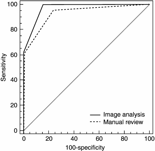
Accurate determination of HER-2 status is critical to identify patients for whom trastuzumab treatment will be of benefit. Although the recommended primary method of evaluation is immunohistochemistry, numerous reports of variability in interpretation have raised uncertainty about the reliability of results. Recent guidelines have suggested that image analysis could be an effective tool for achieving consistent interpretation, and this study aimed to assess whether this technology has potential as a diagnostic support tool.

METHODS AND RESULTS

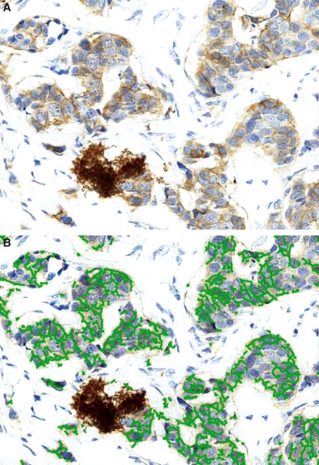
Across a cohort of 275 cases, image analysis could accurately classify HER-2 status, with 91% agreement between computer-aided classification and the pathology review. Assessment of the continuity of membranous immunoreactivity in addition to intensity of reactivity was critical to distinguish between negative and equivocal cases and enabled image analysis to report a lower referral rate of cases for confirmatory fluorescence in situ hybridization (FISH) testing. An excellent concordance rate of 95% was observed between FISH and the automated review across 136 informative cases.

CONCLUSIONS

This study has validated that image analysis can robustly and accurately evaluate HER-2 status in immunohistochemically stained tissue. Based on these findings, image analysis has great potential as a diagnostic support tool for pathologists and biomedical scientists, and may significantly improve the standardization of HER-2 testing by providing a quantitative reference method for interpretation.

摘要

目的

准确确定 HER-2 状态对于识别将从曲妥珠单抗治疗中获益的患者至关重要。虽然推荐的主要评估方法是免疫组织化学,但许多关于解释变异性的报告引起了对结果可靠性的不确定性。最近的指南建议图像分析可能是实现一致解释的有效工具,本研究旨在评估该技术是否有可能成为诊断支持工具。

方法和结果

在 275 例病例的队列中,图像分析可以准确地分类 HER-2 状态,计算机辅助分类与病理审查之间的一致性为 91%。评估膜免疫反应的连续性以及反应强度对于区分阴性和不确定病例至关重要,使图像分析能够报告用于确认荧光原位杂交(FISH)检测的病例的转诊率较低。在 136 例有意义的病例中,FISH 与自动审查之间观察到极好的一致性,达到 95%。

结论

本研究验证了图像分析可以稳健且准确地评估免疫组织化学染色组织中的 HER-2 状态。基于这些发现,图像分析具有作为病理学家和生物医学科学家的诊断支持工具的巨大潜力,并且通过提供用于解释的定量参考方法,可能会显著改善 HER-2 测试的标准化。

https://cdn.ncbi.nlm.nih.gov/pmc/blobs/37ae/2916223/deaab6bf4103/his0057-0027-f6.jpg
https://cdn.ncbi.nlm.nih.gov/pmc/blobs/37ae/2916223/a3ba648cfd50/his0057-0027-f1.jpg
https://cdn.ncbi.nlm.nih.gov/pmc/blobs/37ae/2916223/0397e825680a/his0057-0027-f2.jpg
https://cdn.ncbi.nlm.nih.gov/pmc/blobs/37ae/2916223/9b2330495d90/his0057-0027-f3.jpg
https://cdn.ncbi.nlm.nih.gov/pmc/blobs/37ae/2916223/51951c58e3f6/his0057-0027-f4.jpg
https://cdn.ncbi.nlm.nih.gov/pmc/blobs/37ae/2916223/36bac3667d18/his0057-0027-f5.jpg
https://cdn.ncbi.nlm.nih.gov/pmc/blobs/37ae/2916223/deaab6bf4103/his0057-0027-f6.jpg

相似文献

1
Image analysis as an adjunct to manual HER-2 immunohistochemical review: a diagnostic tool to standardize interpretation.图像分析作为人 HER-2 免疫组化复查的辅助手段:一种标准化解读的诊断工具。
Histopathology. 2010 Jul;57(1):27-38. doi: 10.1111/j.1365-2559.2010.03577.x. Epub 2010 Jun 24.
2
HER-2 status determination in breast carcinomas. A practical approach.乳腺癌中HER-2状态的判定:一种实用方法
Histol Histopathol. 2006 Mar;21(3):227-36. doi: 10.14670/HH-21.227.
3
Achieving 95% cross-methodological concordance in HER2 testing: causes and implications of discordant cases.实现 HER2 检测方法间 95%的一致性:不一致病例的原因及意义。
Am J Clin Pathol. 2010 Aug;134(2):284-92. doi: 10.1309/AJCPUQB18XZOHHBJ.
4
HER-2 testing in breast cancer using parallel tissue-based methods.使用基于组织的平行方法检测乳腺癌中的HER-2
JAMA. 2004 Apr 28;291(16):1972-7. doi: 10.1001/jama.291.16.1972.
5
Computer-assisted assessment of the human epidermal growth factor receptor 2 immunohistochemical assay in imaged histologic sections using a membrane isolation algorithm and quantitative analysis of positive controls.使用膜分离算法和阳性对照的定量分析对成像组织学切片中的人表皮生长因子受体2免疫组织化学检测进行计算机辅助评估。
BMC Med Imaging. 2008 Jun 5;8:11. doi: 10.1186/1471-2342-8-11.
6
[HER2 testing in breast cancer].[乳腺癌中的人表皮生长因子受体2检测]
Nihon Rinsho. 2007 Jun 28;65 Suppl 6:275-80.
7
Enhanced accuracy and reliability of HER-2/neu immunohistochemical scoring using digital microscopy.使用数字显微镜提高HER-2/neu免疫组织化学评分的准确性和可靠性。
Am J Clin Pathol. 2004 May;121(5):620-30. doi: 10.1309/Y73U-8X72-B68T-MGH5.
8
Recent trends of HER-2 testing and trastuzumab therapy for breast cancer.乳腺癌HER-2检测及曲妥珠单抗治疗的近期趋势
Breast Cancer. 2009;16(4):284-7. doi: 10.1007/s12282-009-0159-z. Epub 2009 Aug 6.
9
Quantitative evaluation of HER-2/neu status in breast cancer by fluorescence in situ hybridization and by immunohistochemistry with image analysis.通过荧光原位杂交和图像分析免疫组织化学对乳腺癌中HER-2/neu状态进行定量评估。
Am J Clin Pathol. 2001 Jun;115(6):814-22. doi: 10.1309/AJ84-50AK-1X1B-1Q4C.
10
Accuracy and precision in HER2/neu testing in breast cancer: are we there yet?乳腺癌HER2/neu检测中的准确性和精密度:我们做到了吗?
Hum Pathol. 2004 Feb;35(2):143-6. doi: 10.1016/j.humpath.2004.01.002.

引用本文的文献

1
Deep learning application in prediction of cancer molecular alterations based on pathological images: a bibliographic analysis via CiteSpace.深度学习在基于病理图像的癌症分子改变预测中的应用:基于 CiteSpace 的文献分析。
J Cancer Res Clin Oncol. 2024 Oct 18;150(10):467. doi: 10.1007/s00432-024-05992-z.
2
Fully Automated Artificial Intelligence Solution for Human Epidermal Growth Factor Receptor 2 Immunohistochemistry Scoring in Breast Cancer: A Multireader Study.全自动人工智能解决方案在乳腺癌人表皮生长因子受体 2 免疫组织化学评分中的应用:一项多读者研究。
JCO Precis Oncol. 2024 Oct;8:e2400353. doi: 10.1200/PO.24.00353. Epub 2024 Oct 11.
3

本文引用的文献

1
Automated image analysis in histopathology: a valuable tool in medical diagnostics.组织病理学中的自动图像分析:医学诊断中的一项重要工具。
Expert Rev Mol Diagn. 2008 Nov;8(6):707-25. doi: 10.1586/14737159.8.6.707.
2
Biomarkers in oncology drug development: rescuers or troublemakers?肿瘤学药物研发中的生物标志物:救星还是麻烦制造者?
Expert Opin Drug Metab Toxicol. 2008 Nov;4(11):1391-402. doi: 10.1517/17425255.4.11.1391.
3
Novel image analysis approach for quantifying expression of nuclear proteins assessed by immunohistochemistry: application to measurement of oestrogen and progesterone receptor levels in breast cancer.
HER2 Intratumoral Heterogeneity in Breast Cancer, an Evolving Concept.
乳腺癌中HER2肿瘤内异质性:一个不断发展的概念
Cancers (Basel). 2023 May 9;15(10):2664. doi: 10.3390/cancers15102664.
4
Progress on deep learning in digital pathology of breast cancer: a narrative review.乳腺癌数字病理学中深度学习的进展:一篇综述
Gland Surg. 2022 Apr;11(4):751-766. doi: 10.21037/gs-22-11.
5
Comparison of manual and automated digital image analysis systems for quantification of cellular protein expression.手动和自动数字图像分析系统在细胞蛋白表达定量中的比较。
Histol Histopathol. 2022 Jun;37(6):527-541. doi: 10.14670/HH-18-434. Epub 2022 Feb 11.
6
Intratumoral Heterogeneity and Immune Response Indicators to Predict Overall Survival in a Retrospective Study of HER2-Borderline (IHC 2+) Breast Cancer Patients.一项关于HER2临界(免疫组化2+)乳腺癌患者的回顾性研究中,肿瘤内异质性和免疫反应指标对总生存期的预测作用
Front Oncol. 2021 Nov 11;11:774088. doi: 10.3389/fonc.2021.774088. eCollection 2021.
7
HER2 Molecular Marker Scoring Using Transfer Learning and Decision Level Fusion.使用迁移学习和决策级融合进行 HER2 分子标志物评分。
J Digit Imaging. 2021 Jun;34(3):667-677. doi: 10.1007/s10278-021-00442-5. Epub 2021 Mar 19.
8
Quantitative Image Analysis for Tissue Biomarker Use: A White Paper From the Digital Pathology Association.用于组织生物标志物的定量图像分析:数字病理协会白皮书
Appl Immunohistochem Mol Morphol. 2021 Aug 1;29(7):479-493. doi: 10.1097/PAI.0000000000000930.
9
A Validation Study of Human Epidermal Growth Factor Receptor 2 Immunohistochemistry Digital Imaging Analysis and its Correlation with Human Epidermal Growth Factor Receptor 2 Fluorescence Hybridization Results in Breast Carcinoma.人表皮生长因子受体2免疫组化数字成像分析及其与乳腺癌中人表皮生长因子受体2荧光原位杂交结果的相关性验证研究
J Pathol Inform. 2020 Feb 4;11:2. doi: 10.4103/jpi.jpi_52_19. eCollection 2020.
10
Quantitative digital imaging analysis of HER2 immunohistochemistry predicts the response to anti-HER2 neoadjuvant chemotherapy in HER2-positive breast carcinoma.HER2 阳性乳腺癌抗 HER2 新辅助化疗反应的定量数字成像分析预测。
Breast Cancer Res Treat. 2020 Apr;180(2):321-329. doi: 10.1007/s10549-020-05546-0. Epub 2020 Jan 30.
用于通过免疫组织化学定量评估核蛋白表达的新型图像分析方法:在乳腺癌雌激素和孕激素受体水平测量中的应用。
Breast Cancer Res. 2008;10(5):R89. doi: 10.1186/bcr2187. Epub 2008 Oct 23.
4
Virtual microscopy as an enabler of automated/quantitative assessment of protein expression in TMAs.虚拟显微镜技术助力组织微阵列中蛋白质表达的自动化/定量评估。
Histochem Cell Biol. 2008 Sep;130(3):447-63. doi: 10.1007/s00418-008-0480-1. Epub 2008 Aug 5.
5
Creation of a digital slide and tissue microarray resource from a multi-institutional predictive toxicology study in the rat: an initial report from the PredTox group.利用大鼠多机构预测毒理学研究创建数字玻片和组织微阵列资源:PredTox 小组的初步报告
Exp Toxicol Pathol. 2008 Aug;60(4-5):235-45. doi: 10.1016/j.etp.2007.12.004. Epub 2008 May 13.
6
HER2 testing in the UK: further update to recommendations.英国的 HER2 检测:建议的进一步更新。
J Clin Pathol. 2008 Jul;61(7):818-24. doi: 10.1136/jcp.2007.054866. Epub 2008 Apr 1.
7
A quality assurance exercise to evaluate the accuracy and reproducibility of chromogenic in situ hybridisation for HER2 analysis in breast cancer.一项用于评估乳腺癌中HER2分析的显色原位杂交准确性和可重复性的质量保证活动。
J Clin Pathol. 2008 Jun;61(6):757-60. doi: 10.1136/jcp.2007.053850. Epub 2008 Mar 6.
8
HER2+ breast cancer: review of biologic relevance and optimal use of diagnostic tools.人表皮生长因子受体2阳性乳腺癌:生物学相关性及诊断工具最佳应用的综述
Am J Clin Pathol. 2008 Feb;129(2):263-73. doi: 10.1309/99AE032R9FM8WND1.
9
Assessment of two automated imaging systems in evaluating estrogen receptor status in breast carcinoma.两种自动成像系统在评估乳腺癌雌激素受体状态中的应用
Appl Immunohistochem Mol Morphol. 2007 Dec;15(4):451-5. doi: 10.1097/PAI.0b013e31802ee998.
10
American Society of Clinical Oncology 2007 update of recommendations for the use of tumor markers in breast cancer.美国临床肿瘤学会2007年乳腺癌肿瘤标志物应用建议更新版。
J Clin Oncol. 2007 Nov 20;25(33):5287-312. doi: 10.1200/JCO.2007.14.2364. Epub 2007 Oct 22.