• 文献检索
  • 文档翻译
  • 深度研究
  • 学术资讯
  • Suppr Zotero 插件Zotero 插件
  • 邀请有礼
  • 套餐&价格
  • 历史记录
应用&插件
Suppr Zotero 插件Zotero 插件浏览器插件Mac 客户端Windows 客户端微信小程序
定价
高级版会员购买积分包购买API积分包
服务
文献检索文档翻译深度研究API 文档MCP 服务
关于我们
关于 Suppr公司介绍联系我们用户协议隐私条款
关注我们

Suppr 超能文献

核心技术专利:CN118964589B侵权必究
粤ICP备2023148730 号-1Suppr @ 2026

文献检索

告别复杂PubMed语法,用中文像聊天一样搜索,搜遍4000万医学文献。AI智能推荐,让科研检索更轻松。

立即免费搜索

文件翻译

保留排版,准确专业,支持PDF/Word/PPT等文件格式,支持 12+语言互译。

免费翻译文档

深度研究

AI帮你快速写综述,25分钟生成高质量综述,智能提取关键信息,辅助科研写作。

立即免费体验

在体内聚焦 T2 加权 MRI 中对海马亚区进行近乎自动的分割。

Nearly automatic segmentation of hippocampal subfields in in vivo focal T2-weighted MRI.

机构信息

Penn Image Computing and Science Laboratory, Department of Radiology, University of Pennsylvania, Philadelphia, PA, USA.

出版信息

Neuroimage. 2010 Dec;53(4):1208-24. doi: 10.1016/j.neuroimage.2010.06.040. Epub 2010 Jun 30.

DOI:10.1016/j.neuroimage.2010.06.040
PMID:20600984
原文链接:https://pmc.ncbi.nlm.nih.gov/articles/PMC2939190/
Abstract

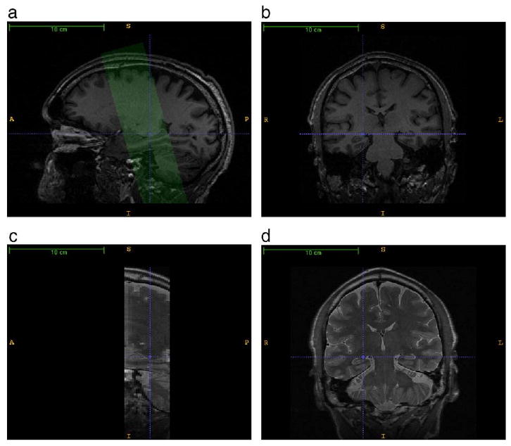
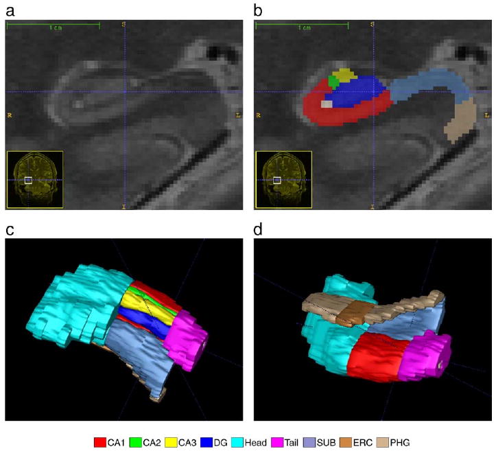
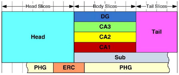
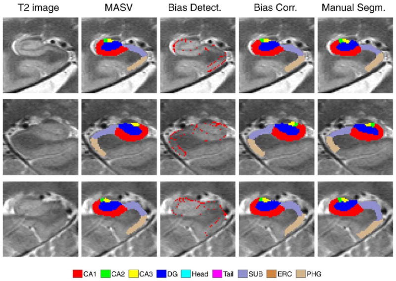
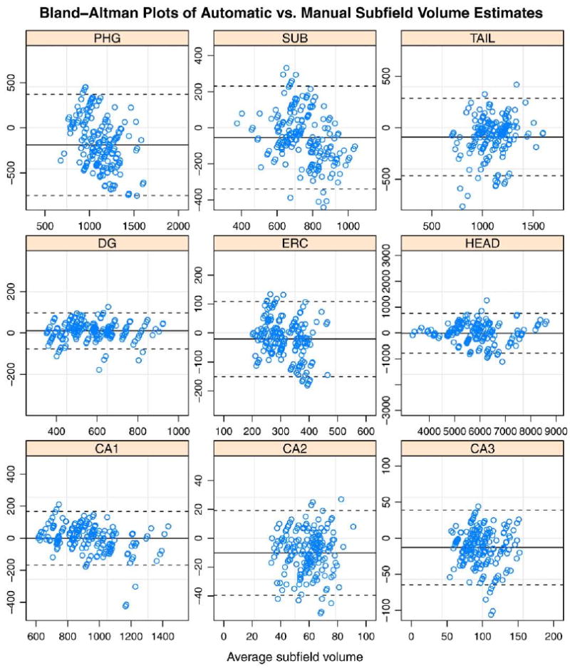
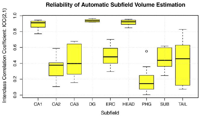
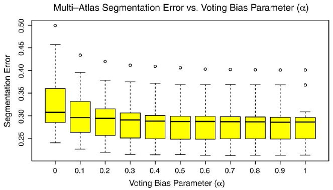
We present and evaluate a new method for automatically labeling the subfields of the hippocampal formation in focal 0.4 × 0.5 × 2.0mm(3) resolution T2-weighted magnetic resonance images that can be acquired in the routine clinical setting with under 5 min scan time. The method combines multi-atlas segmentation, similarity-weighted voting, and a novel learning-based bias correction technique to achieve excellent agreement with manual segmentation. Initial partitioning of MRI slices into hippocampal 'head', 'body' and 'tail' slices is the only input required from the user, necessitated by the nature of the underlying segmentation protocol. Dice overlap between manual and automatic segmentation is above 0.87 for the larger subfields, CA1 and dentate gyrus, and is competitive with the best results for whole-hippocampus segmentation in the literature. Intraclass correlation of volume measurements in CA1 and dentate gyrus is above 0.89. Overlap in smaller hippocampal subfields is lower in magnitude (0.54 for CA2, 0.62 for CA3, 0.77 for subiculum and 0.79 for entorhinal cortex) but comparable to overlap between manual segmentations by trained human raters. These results support the feasibility of subfield-specific hippocampal morphometry in clinical studies of memory and neurodegenerative disease.

摘要

我们提出并评估了一种新方法,用于自动标记海马结构的子字段,这些子字段在常规临床环境中以不到 5 分钟的扫描时间获取,分辨率为 0.4×0.5×2.0mm(3)的 T2 加权磁共振图像。该方法结合了多图谱分割、相似性加权投票和一种新颖的基于学习的偏差校正技术,可实现与手动分割的优异一致性。MRI 切片的初始分区为海马的“头”、“体”和“尾”切片,这是由于底层分割协议的性质,是用户唯一需要的输入。手动和自动分割之间的 Dice 重叠大于 0.87,适用于较大的子字段 CA1 和齿状回,与文献中整个海马体分割的最佳结果相当。在 CA1 和齿状回的体积测量中,组内相关系数大于 0.89。较小的海马子字段的重叠程度较低(CA2 为 0.54,CA3 为 0.62,下托为 0.77,内嗅皮层为 0.79),但与经过训练的人类评估者的手动分割之间的重叠相当。这些结果支持在记忆和神经退行性疾病的临床研究中进行特定子字段的海马形态计量学的可行性。

https://cdn.ncbi.nlm.nih.gov/pmc/blobs/dd21/2939190/953b5b6f87b2/nihms220223f12.jpg
https://cdn.ncbi.nlm.nih.gov/pmc/blobs/dd21/2939190/492a53fcfda3/nihms220223f1.jpg
https://cdn.ncbi.nlm.nih.gov/pmc/blobs/dd21/2939190/5e92732f0c7e/nihms220223f2.jpg
https://cdn.ncbi.nlm.nih.gov/pmc/blobs/dd21/2939190/fae8a211927b/nihms220223f3.jpg
https://cdn.ncbi.nlm.nih.gov/pmc/blobs/dd21/2939190/746f68a894ab/nihms220223f4.jpg
https://cdn.ncbi.nlm.nih.gov/pmc/blobs/dd21/2939190/8ebd9df029d3/nihms220223f5.jpg
https://cdn.ncbi.nlm.nih.gov/pmc/blobs/dd21/2939190/776c9ab868eb/nihms220223f6.jpg
https://cdn.ncbi.nlm.nih.gov/pmc/blobs/dd21/2939190/2bb500f3948d/nihms220223f7.jpg
https://cdn.ncbi.nlm.nih.gov/pmc/blobs/dd21/2939190/9b5542126c53/nihms220223f8.jpg
https://cdn.ncbi.nlm.nih.gov/pmc/blobs/dd21/2939190/9f29ed9a568e/nihms220223f9.jpg
https://cdn.ncbi.nlm.nih.gov/pmc/blobs/dd21/2939190/02ef602d2c09/nihms220223f10.jpg
https://cdn.ncbi.nlm.nih.gov/pmc/blobs/dd21/2939190/ec06732c3891/nihms220223f11.jpg
https://cdn.ncbi.nlm.nih.gov/pmc/blobs/dd21/2939190/953b5b6f87b2/nihms220223f12.jpg

相似文献

1
Nearly automatic segmentation of hippocampal subfields in in vivo focal T2-weighted MRI.在体内聚焦 T2 加权 MRI 中对海马亚区进行近乎自动的分割。
Neuroimage. 2010 Dec;53(4):1208-24. doi: 10.1016/j.neuroimage.2010.06.040. Epub 2010 Jun 30.
2
Automated Hippocampal Subfield Segmentation at 7T MRI.7T磁共振成像下的海马亚区自动分割
AJNR Am J Neuroradiol. 2016 Jun;37(6):1050-7. doi: 10.3174/ajnr.A4659. Epub 2016 Feb 4.
3
Multi-atlas segmentation of the whole hippocampus and subfields using multiple automatically generated templates.使用多个自动生成的模板对整个海马体及其子区进行多图谱分割。
Neuroimage. 2014 Nov 1;101:494-512. doi: 10.1016/j.neuroimage.2014.04.054. Epub 2014 Apr 29.
4
Automated volumetry and regional thickness analysis of hippocampal subfields and medial temporal cortical structures in mild cognitive impairment.轻度认知障碍中海马亚区及内侧颞叶皮质结构的自动容积测量和区域厚度分析
Hum Brain Mapp. 2015 Jan;36(1):258-87. doi: 10.1002/hbm.22627. Epub 2014 Sep 2.
5
Development of a histologically validated segmentation protocol for the hippocampal body.开发一种基于组织学验证的海马体分割协议。
Neuroimage. 2017 Aug 15;157:219-232. doi: 10.1016/j.neuroimage.2017.06.008. Epub 2017 Jun 3.
6
A protocol for manual segmentation of medial temporal lobe subregions in 7 Tesla MRI.一种在7特斯拉磁共振成像中手动分割内侧颞叶亚区域的方案。
Neuroimage Clin. 2017 May 26;15:466-482. doi: 10.1016/j.nicl.2017.05.022. eCollection 2017.
7
Subfields of the hippocampal formation at 7 T MRI: in vivo volumetric assessment.7T MRI 下海马结构的亚区:活体容积评估。
Neuroimage. 2012 Jul 16;61(4):1043-9. doi: 10.1016/j.neuroimage.2012.03.023. Epub 2012 Mar 14.
8
A novel in vivo atlas of human hippocampal subfields using high-resolution 3 T magnetic resonance imaging.利用高分辨率 3T 磁共振成像构建人类海马亚区的新型活体图谱。
Neuroimage. 2013 Jul 1;74:254-65. doi: 10.1016/j.neuroimage.2013.02.003. Epub 2013 Feb 13.
9
Curved multiplanar reformatting allows the accurate histological delineation of hippocampal subfields.曲面多平面重建可实现对海马亚区的准确组织学描绘。
Hippocampus. 2024 Nov;34(11):625-632. doi: 10.1002/hipo.23637. Epub 2024 Sep 11.
10
Quantitative comparison of 21 protocols for labeling hippocampal subfields and parahippocampal subregions in in vivo MRI: towards a harmonized segmentation protocol.21种用于在活体磁共振成像中标记海马亚区和海马旁回亚区域的方案的定量比较:迈向统一的分割方案
Neuroimage. 2015 May 1;111:526-41. doi: 10.1016/j.neuroimage.2015.01.004. Epub 2015 Jan 14.

引用本文的文献

1
Developing an Anatomically Valid Segmentation Protocol for Anterior Regions of the Medial Temporal Lobe for Neurodegenerative Diseases.为神经退行性疾病开发一种用于内侧颞叶前部区域的解剖学上有效的分割方案。
Hippocampus. 2025 Sep;35(5):e70027. doi: 10.1002/hipo.70027.
2
Harmonized Protocol for Subfield Segmentation in the Hippocampal Body on High-Resolution MRI from the Hippocampal Subfields Group (HSG).海马体亚区组(HSG)高分辨率MRI上海马体中部亚区分割的统一协议。
bioRxiv. 2025 May 4:2025.04.29.651039. doi: 10.1101/2025.04.29.651039.
3
Developing an anatomically valid segmentation protocol for anterior regions of the medial temporal lobe for neurodegenerative diseases.

本文引用的文献

1
Towards accurate, automatic segmentation of the hippocampus and amygdala from MRI.迈向从磁共振成像中准确、自动分割海马体和杏仁核。
Med Image Comput Comput Assist Interv. 2009;12(Pt 2):592-600. doi: 10.1007/978-3-642-04271-3_72.
2
High-resolution structural and functional MRI of hippocampal CA3 and dentate gyrus in patients with amnestic Mild Cognitive Impairment.遗忘型轻度认知障碍患者海马 CA3 和齿状回的高分辨率结构和功能 MRI。
Neuroimage. 2010 Jul 1;51(3):1242-52. doi: 10.1016/j.neuroimage.2010.03.040. Epub 2010 Mar 23.
3
Magnetic resonance imaging of hippocampal subfields in posttraumatic stress disorder.
为神经退行性疾病开发一种用于内侧颞叶前部区域的解剖学上有效的分割方案。
bioRxiv. 2025 Feb 13:2025.02.11.637506. doi: 10.1101/2025.02.11.637506.
4
A (sub)field guide to quality control in hippocampal subfield segmentation on high-resolution T-weighted MRI.高分辨率 T1 加权 MRI 中海马亚区分割的质量控制 (子) 领域指南。
Hum Brain Mapp. 2024 Oct 15;45(15):e70004. doi: 10.1002/hbm.70004.
5
Morphometry of medial temporal lobe subregions using high-resolution T2-weighted MRI in ADNI3: Why, how, and what's next?ADNI3 中使用高分辨率 T2 加权 MRI 测量内侧颞叶亚区的形态计量学:为什么、如何以及下一步是什么?
Alzheimers Dement. 2024 Nov;20(11):8113-8128. doi: 10.1002/alz.14161. Epub 2024 Sep 16.
6
Amygdala atrophies in specific subnuclei in preclinical Alzheimer's disease.在临床前阿尔茨海默病中,杏仁核特定亚核发生萎缩。
Alzheimers Dement. 2024 Oct;20(10):7205-7219. doi: 10.1002/alz.14235. Epub 2024 Sep 10.
7
DSnet: a new dual-branch network for hippocampus subfield segmentation.DSnet:一种新的双分支网络,用于海马亚区分割。
Sci Rep. 2024 Jul 3;14(1):15317. doi: 10.1038/s41598-024-66415-0.
8
Structural Neuroimaging of Hippocampus and Amygdala Subregions in Posttraumatic Stress Disorder: A Scoping Review.创伤后应激障碍中海马体和杏仁核亚区域的结构神经影像学:一项范围综述
Biol Psychiatry Glob Open Sci. 2023 Jul 13;4(1):120-134. doi: 10.1016/j.bpsgos.2023.07.001. eCollection 2024 Jan.
9
A (Sub)field Guide to Quality Control in Hippocampal Subfield Segmentation on Highresolution T-weighted MRI.高分辨率T加权MRI海马亚区分割质量控制(子)领域指南
bioRxiv. 2023 Dec 1:2023.11.29.568895. doi: 10.1101/2023.11.29.568895.
10
Examining the efficacy of a cardio-dance intervention on brain health and the moderating role of ABCA7 in older African Americans: a protocol for a randomized controlled trial.探讨有氧舞蹈干预对老年非裔美国人脑健康的疗效及ABCA7的调节作用:一项随机对照试验方案
Front Aging Neurosci. 2023 Nov 21;15:1266423. doi: 10.3389/fnagi.2023.1266423. eCollection 2023.
创伤后应激障碍中海马亚区的磁共振成像
Arch Gen Psychiatry. 2010 Mar;67(3):296-303. doi: 10.1001/archgenpsychiatry.2009.205.
4
The optimal template effect in hippocampus studies of diseased populations.疾病人群海马体研究中的最佳模板效应。
Neuroimage. 2010 Feb 1;49(3):2457-66. doi: 10.1016/j.neuroimage.2009.09.062. Epub 2009 Oct 8.
5
In vivo quantification of hippocampal subfields using 4.7 T fast spin echo imaging.使用 4.7T 快速自旋回波成像进行海马亚区的体内定量分析。
Neuroimage. 2010 Jan 15;49(2):1224-30. doi: 10.1016/j.neuroimage.2009.09.042. Epub 2009 Sep 26.
6
Human hippocampal CA1 involvement during allocentric encoding of spatial information.人类海马体CA1在空间信息的以自我为中心编码过程中的参与。
J Neurosci. 2009 Aug 26;29(34):10512-9. doi: 10.1523/JNEUROSCI.0621-09.2009.
7
Selective effect of age, Apo e4, and Alzheimer's disease on hippocampal subfields.年龄、载脂蛋白E4和阿尔茨海默病对海马亚区的选择性影响。
Hippocampus. 2009 Jun;19(6):558-64. doi: 10.1002/hipo.20614.
8
Automated segmentation of hippocampal subfields from ultra-high resolution in vivo MRI.基于超高分辨率活体磁共振成像的海马亚区自动分割
Hippocampus. 2009 Jun;19(6):549-57. doi: 10.1002/hipo.20615.
9
Subfield atrophy pattern in temporal lobe epilepsy with and without mesial sclerosis detected by high-resolution MRI at 4 Tesla: preliminary results.通过4特斯拉高分辨率磁共振成像检测的伴有和不伴有内侧颞叶硬化的颞叶癫痫的脑叶亚区萎缩模式:初步结果
Epilepsia. 2009 Jun;50(6):1474-83. doi: 10.1111/j.1528-1167.2009.02010.x. Epub 2009 Mar 12.
10
MRI of hippocampal volume loss in early Alzheimer's disease in relation to ApoE genotype and biomarkers.早期阿尔茨海默病中海马体积丢失与载脂蛋白E基因型及生物标志物关系的磁共振成像研究
Brain. 2009 Apr;132(Pt 4):1067-77. doi: 10.1093/brain/awp007. Epub 2009 Feb 27.