• 文献检索
  • 文档翻译
  • 深度研究
  • 学术资讯
  • Suppr Zotero 插件Zotero 插件
  • 邀请有礼
  • 套餐&价格
  • 历史记录
应用&插件
Suppr Zotero 插件Zotero 插件浏览器插件Mac 客户端Windows 客户端微信小程序
定价
高级版会员购买积分包购买API积分包
服务
文献检索文档翻译深度研究API 文档MCP 服务
关于我们
关于 Suppr公司介绍联系我们用户协议隐私条款
关注我们

Suppr 超能文献

核心技术专利:CN118964589B侵权必究
粤ICP备2023148730 号-1Suppr @ 2026

文献检索

告别复杂PubMed语法,用中文像聊天一样搜索,搜遍4000万医学文献。AI智能推荐,让科研检索更轻松。

立即免费搜索

文件翻译

保留排版,准确专业,支持PDF/Word/PPT等文件格式,支持 12+语言互译。

免费翻译文档

深度研究

AI帮你快速写综述,25分钟生成高质量综述,智能提取关键信息,辅助科研写作。

立即免费体验

一种在7特斯拉磁共振成像中手动分割内侧颞叶亚区域的方案。

A protocol for manual segmentation of medial temporal lobe subregions in 7 Tesla MRI.

作者信息

Berron D, Vieweg P, Hochkeppler A, Pluta J B, Ding S-L, Maass A, Luther A, Xie L, Das S R, Wolk D A, Wolbers T, Yushkevich P A, Düzel E, Wisse L E M

机构信息

Institute of Cognitive Neurology and Dementia Research, Otto-von-Guericke-University Magdeburg, 39120 Magdeburg, Germany; German Center for Neurodegenerative Diseases (DZNE), Site Magdeburg, 39120 Magdeburg, Germany.

German Center for Neurodegenerative Diseases (DZNE), Site Magdeburg, 39120 Magdeburg, Germany.

出版信息

Neuroimage Clin. 2017 May 26;15:466-482. doi: 10.1016/j.nicl.2017.05.022. eCollection 2017.

DOI:10.1016/j.nicl.2017.05.022
PMID:28652965
原文链接:
https://pmc.ncbi.nlm.nih.gov/articles/PMC5476466/
Abstract

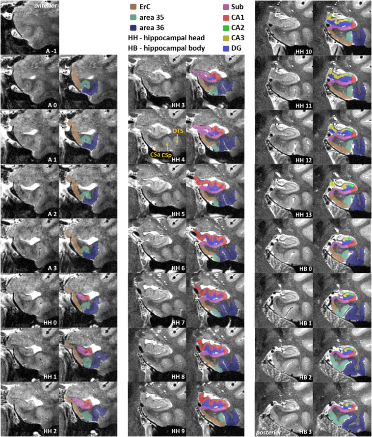
Recent advances in MRI and increasing knowledge on the characterization and anatomical variability of medial temporal lobe (MTL) anatomy have paved the way for more specific subdivisions of the MTL in humans. In addition, recent studies suggest that early changes in many neurodegenerative and neuropsychiatric diseases are better detected in smaller subregions of the MTL rather than with whole structure analyses. Here, we developed a new protocol using 7 Tesla (T) MRI incorporating novel anatomical findings for the manual segmentation of entorhinal cortex (ErC), perirhinal cortex (PrC; divided into area 35 and 36), parahippocampal cortex (PhC), and hippocampus; which includes the subfields subiculum (Sub), CA1, CA2, as well as CA3 and dentate gyrus (DG) which are separated by the endfolial pathway covering most of the long axis of the hippocampus. We provide detailed instructions alongside slice-by-slice segmentations to ease learning for the untrained but also more experienced raters. Twenty-two subjects were scanned (19-32 yrs, mean age = 26 years, 12 females) with a turbo spin echo (TSE) T2-weighted MRI sequence with high-resolution oblique coronal slices oriented orthogonal to the long axis of the hippocampus (in-plane resolution 0.44 × 0.44 mm) and 1.0 mm slice thickness. The scans were manually delineated by two experienced raters, to assess intra- and inter-rater reliability. The Dice Similarity Index (DSI) was above 0.78 for all regions and the Intraclass Correlation Coefficients (ICC) were between 0.76 to 0.99 both for intra- and inter-rater reliability. In conclusion, this study presents a fine-grained and comprehensive segmentation protocol for MTL structures at 7 T MRI that closely follows recent knowledge from anatomical studies. More specific subdivisions (e.g. area 35 and 36 in PrC, and the separation of DG and CA3) may pave the way for more precise delineations thereby enabling the detection of early volumetric changes in dementia and neuropsychiatric diseases.

摘要

磁共振成像(MRI)的最新进展以及对内侧颞叶(MTL)解剖结构特征和解剖变异性的认识不断增加,为人类MTL更具体的细分奠定了基础。此外,最近的研究表明,在许多神经退行性疾病和神经精神疾病中,早期变化在MTL的较小亚区域中比在整体结构分析中更容易被检测到。在此,我们开发了一种新的方案,使用7特斯拉(T)MRI,并结合了新的解剖学发现,用于手动分割内嗅皮质(ErC)、嗅周皮质(PrC;分为35区和36区)、海马旁皮质(PhC)和海马体;其中海马体包括亚区下托(Sub)、CA1、CA2以及CA3和齿状回(DG),它们由覆盖海马体长轴大部分的终叶通路分隔开。我们提供了详细的说明以及逐片分割,以便于未受过训练但经验更丰富的评分者学习。对22名受试者(年龄19 - 32岁,平均年龄 = 26岁,12名女性)进行了扫描,采用快速自旋回波(TSE)T2加权MRI序列,具有与海马体长轴正交的高分辨率斜冠状切片(平面分辨率0.44×0.44毫米),切片厚度为1.0毫米。扫描由两名经验丰富的评分者进行手动描绘,以评估评分者内和评分者间的可靠性。所有区域的骰子相似性指数(DSI)均高于0.78,评分者内和评分者间可靠性的组内相关系数(ICC)在0.76至0.99之间。总之,本研究提出了一种在7T MRI下对MTL结构进行细粒度和全面的分割方案,该方案紧密遵循解剖学研究的最新知识。更具体的细分(例如PrC中的35区和36区,以及DG和CA3的分离)可能为更精确的描绘铺平道路,从而能够检测痴呆和神经精神疾病中的早期体积变化。

https://cdn.ncbi.nlm.nih.gov/pmc/blobs/60f8/5476466/ba554a0e9bcd/gr9.jpg
https://cdn.ncbi.nlm.nih.gov/pmc/blobs/60f8/5476466/00c2e45e0ff7/gr1.jpg
https://cdn.ncbi.nlm.nih.gov/pmc/blobs/60f8/5476466/2c006980b5bc/gr2.jpg
https://cdn.ncbi.nlm.nih.gov/pmc/blobs/60f8/5476466/974e97e17799/gr3.jpg
https://cdn.ncbi.nlm.nih.gov/pmc/blobs/60f8/5476466/7ab66c5def97/gr4.jpg
https://cdn.ncbi.nlm.nih.gov/pmc/blobs/60f8/5476466/3e75361568f6/gr5.jpg
https://cdn.ncbi.nlm.nih.gov/pmc/blobs/60f8/5476466/cc2c28eefcc2/gr6.jpg
https://cdn.ncbi.nlm.nih.gov/pmc/blobs/60f8/5476466/c330aa23832a/gr7.jpg
https://cdn.ncbi.nlm.nih.gov/pmc/blobs/60f8/5476466/7320903d9721/gr8.jpg
https://cdn.ncbi.nlm.nih.gov/pmc/blobs/60f8/5476466/ba554a0e9bcd/gr9.jpg

相似文献

1
A protocol for manual segmentation of medial temporal lobe subregions in 7 Tesla MRI.一种在7特斯拉磁共振成像中手动分割内侧颞叶亚区域的方案。
Neuroimage Clin. 2017 May 26;15:466-482. doi: 10.1016/j.nicl.2017.05.022. eCollection 2017.
2
Automated Hippocampal Subfield Segmentation at 7T MRI.7T磁共振成像下的海马亚区自动分割
AJNR Am J Neuroradiol. 2016 Jun;37(6):1050-7. doi: 10.3174/ajnr.A4659. Epub 2016 Feb 4.
3
Downstream effects of polypathology on neurodegeneration of medial temporal lobe subregions.多病理学对内侧颞叶亚区神经退行性变的下游影响。
Acta Neuropathol Commun. 2021 Jul 21;9(1):128. doi: 10.1186/s40478-021-01225-3.
4
Quantitative comparison of 21 protocols for labeling hippocampal subfields and parahippocampal subregions in in vivo MRI: towards a harmonized segmentation protocol.21种用于在活体磁共振成像中标记海马亚区和海马旁回亚区域的方案的定量比较:迈向统一的分割方案
Neuroimage. 2015 May 1;111:526-41. doi: 10.1016/j.neuroimage.2015.01.004. Epub 2015 Jan 14.
5
Volumetric analysis of medial temporal lobe subregions in developmental amnesia using high-resolution magnetic resonance imaging.使用高分辨率磁共振成像对发育性遗忘症内侧颞叶亚区进行容积分析。
Hippocampus. 2013 Oct;23(10):855-60. doi: 10.1002/hipo.22153. Epub 2013 Jul 5.
6
Intrinsic functional connectivity of the human medial temporal lobe suggests a distinction between adjacent MTL cortices and hippocampus.人类内侧颞叶的内在功能连接表明相邻的 MTL 皮质和海马之间存在区别。
Hippocampus. 2012 Dec;22(12):2290-302. doi: 10.1002/hipo.22047. Epub 2012 Jun 27.
7
Automated volumetry and regional thickness analysis of hippocampal subfields and medial temporal cortical structures in mild cognitive impairment.轻度认知障碍中海马亚区及内侧颞叶皮质结构的自动容积测量和区域厚度分析
Hum Brain Mapp. 2015 Jan;36(1):258-87. doi: 10.1002/hbm.22627. Epub 2014 Sep 2.
8
Subfields of the hippocampal formation at 7 T MRI: in vivo volumetric assessment.7T MRI 下海马结构的亚区:活体容积评估。
Neuroimage. 2012 Jul 16;61(4):1043-9. doi: 10.1016/j.neuroimage.2012.03.023. Epub 2012 Mar 14.
9
Hippocampal Stratum Radiatum, Lacunosum, and Moleculare Sparing in Mild Cognitive Impairment.轻度认知障碍中海马放射层、腔隙层和分子层的保留。
J Alzheimers Dis. 2018;61(1):415-424. doi: 10.3233/JAD-170344.
10
Functional connectivity based parcellation of the human medial temporal lobe.基于功能连接的人类内侧颞叶脑区划分
Neurobiol Learn Mem. 2016 Oct;134 Pt A(Pt A):123-134. doi: 10.1016/j.nlm.2016.01.005. Epub 2016 Jan 19.

引用本文的文献

1
Representational similarity analysis reveals cue-independent spatial representations for landmarks and self-motion cues in human retrosplenial cortex.表征相似性分析揭示了人类压后皮质中地标和自我运动线索的与线索无关的空间表征。
Imaging Neurosci (Camb). 2025 Mar 24;3. doi: 10.1162/imag_a_00516. eCollection 2025.
2
Developing an Anatomically Valid Segmentation Protocol for Anterior Regions of the Medial Temporal Lobe for Neurodegenerative Diseases.为神经退行性疾病开发一种用于内侧颞叶前部区域的解剖学上有效的分割方案。
Hippocampus. 2025 Sep;35(5):e70027. doi: 10.1002/hipo.70027.
3
Harmonized Protocol for Subfield Segmentation in the Hippocampal Body on High-Resolution MRI from the Hippocampal Subfields Group (HSG).

本文引用的文献

1
Accounting for the Confound of Meninges in Segmenting Entorhinal and Perirhinal Cortices in T1-Weighted MRI.在T1加权磁共振成像中分割内嗅皮质和嗅周皮质时对脑膜混杂因素的考量
Med Image Comput Comput Assist Interv. 2016 Oct;9901:564-571. doi: 10.1007/978-3-319-46723-8_65. Epub 2016 Oct 2.
2
A harmonized segmentation protocol for hippocampal and parahippocampal subregions: Why do we need one and what are the key goals?一种用于海马体和海马旁回亚区域的统一分割方案:我们为何需要它以及关键目标是什么?
Hippocampus. 2017 Jan;27(1):3-11. doi: 10.1002/hipo.22671. Epub 2016 Nov 15.
3
Medial temporal lobe subregional morphometry using high resolution MRI in Alzheimer's disease.
海马体亚区组(HSG)高分辨率MRI上海马体中部亚区分割的统一协议。
bioRxiv. 2025 May 4:2025.04.29.651039. doi: 10.1101/2025.04.29.651039.
4
Reading specific memories from human neurons before and after sleep.睡眠前后从人类神经元中读取特定记忆。
bioRxiv. 2025 Jul 4:2025.07.01.662486. doi: 10.1101/2025.07.01.662486.
5
Controlling sharpness, SNR, and specific absorption rate for 3D fast-spin echo at 7T by end-to-end learning.通过端到端学习控制7T下3D快速自旋回波的锐利度、信噪比和比吸收率。
Magn Reson Med. 2025 Sep;94(3):1026-1043. doi: 10.1002/mrm.30533. Epub 2025 May 23.
6
Effects of Early Midlife Ovarian Removal on Medial Temporal Lobe Gray Matter Volume and Recognition Memory.中年早期卵巢切除对内侧颞叶灰质体积和识别记忆的影响。
Hippocampus. 2025 May;35(3):e70012. doi: 10.1002/hipo.70012.
7
Developing an anatomically valid segmentation protocol for anterior regions of the medial temporal lobe for neurodegenerative diseases.为神经退行性疾病开发一种用于内侧颞叶前部区域的解剖学上有效的分割方案。
bioRxiv. 2025 Feb 13:2025.02.11.637506. doi: 10.1101/2025.02.11.637506.
8
A paired dataset of multi-modal MRI at 3 Tesla and 7 Tesla with manual hippocampal subfield segmentations.一个包含3特斯拉和7特斯拉多模态磁共振成像以及手动海马亚区分割的配对数据集。
Sci Data. 2025 Feb 13;12(1):260. doi: 10.1038/s41597-025-04586-9.
9
Medial temporal lobe atrophy patterns in early-versus late-onset amnestic Alzheimer's disease.早期和晚期发病遗忘型阿尔茨海默病的内侧颞叶萎缩模式。
Alzheimers Res Ther. 2024 Sep 16;16(1):204. doi: 10.1186/s13195-024-01571-z.
10
Amygdala atrophies in specific subnuclei in preclinical Alzheimer's disease.在临床前阿尔茨海默病中,杏仁核特定亚核发生萎缩。
Alzheimers Dement. 2024 Oct;20(10):7205-7219. doi: 10.1002/alz.14235. Epub 2024 Sep 10.
使用高分辨率磁共振成像对阿尔茨海默病进行内侧颞叶亚区域形态测量
Neurobiol Aging. 2017 Jan;49:204-213. doi: 10.1016/j.neurobiolaging.2016.09.011. Epub 2016 Sep 30.
4
Multi-template analysis of human perirhinal cortex in brain MRI: Explicitly accounting for anatomical variability.脑磁共振成像中人类嗅周皮质的多模板分析:明确考虑解剖变异性。
Neuroimage. 2017 Jan 1;144(Pt A):183-202. doi: 10.1016/j.neuroimage.2016.09.070. Epub 2016 Oct 1.
5
The Human Dentate Gyrus Plays a Necessary Role in Discriminating New Memories.人类齿状回在区分新记忆方面发挥着必要作用。
Curr Biol. 2016 Oct 10;26(19):2629-2634. doi: 10.1016/j.cub.2016.07.081. Epub 2016 Sep 22.
6
Comparison of In Vivo and Ex Vivo MRI of the Human Hippocampal Formation in the Same Subjects.同一受试者体内和离体 MRI 对人类海马结构的比较。
Cereb Cortex. 2017 Nov 1;27(11):5185-5196. doi: 10.1093/cercor/bhw299.
7
Strong Evidence for Pattern Separation in Human Dentate Gyrus.人类齿状回中模式分离的有力证据。
J Neurosci. 2016 Jul 20;36(29):7569-79. doi: 10.1523/JNEUROSCI.0518-16.2016.
8
Comprehensive cellular-resolution atlas of the adult human brain.成年人类大脑的全细胞分辨率图谱。
J Comp Neurol. 2016 Nov 1;524(16):3127-481. doi: 10.1002/cne.24080.
9
Automated Hippocampal Subfield Segmentation at 7T MRI.7T磁共振成像下的海马亚区自动分割
AJNR Am J Neuroradiol. 2016 Jun;37(6):1050-7. doi: 10.3174/ajnr.A4659. Epub 2016 Feb 4.
10
Age differences in hippocampal subfield volumes from childhood to late adulthood.从童年到成年晚期海马体亚区体积的年龄差异。
Hippocampus. 2016 Feb;26(2):220-8. doi: 10.1002/hipo.22517. Epub 2015 Sep 4.