• 文献检索
  • 文档翻译
  • 深度研究
  • 学术资讯
  • Suppr Zotero 插件Zotero 插件
  • 邀请有礼
  • 套餐&价格
  • 历史记录
应用&插件
Suppr Zotero 插件Zotero 插件浏览器插件Mac 客户端Windows 客户端微信小程序
定价
高级版会员购买积分包购买API积分包
服务
文献检索文档翻译深度研究API 文档MCP 服务
关于我们
关于 Suppr公司介绍联系我们用户协议隐私条款
关注我们

Suppr 超能文献

核心技术专利:CN118964589B侵权必究
粤ICP备2023148730 号-1Suppr @ 2026

文献检索

告别复杂PubMed语法,用中文像聊天一样搜索,搜遍4000万医学文献。AI智能推荐,让科研检索更轻松。

立即免费搜索

文件翻译

保留排版,准确专业,支持PDF/Word/PPT等文件格式,支持 12+语言互译。

免费翻译文档

深度研究

AI帮你快速写综述,25分钟生成高质量综述,智能提取关键信息,辅助科研写作。

立即免费体验

扩展形态学处理:一种从微观图像中自动检测生物标记物的实用方法。

Extended morphological processing: a practical method for automatic spot detection of biological markers from microscopic images.

机构信息

Department of Ultrastructural Research, National Institute of Neuroscience, National Center of Neurology and Psychiatry, Ogawahigashi-cho 4-1-1, Kodaira, Tokyo 187-8502, Japan.

出版信息

BMC Bioinformatics. 2010 Jul 8;11:373. doi: 10.1186/1471-2105-11-373.

DOI:10.1186/1471-2105-11-373
PMID:20615231
原文链接:https://pmc.ncbi.nlm.nih.gov/articles/PMC2914730/
Abstract

BACKGROUND

A reliable extraction technique for resolving multiple spots in light or electron microscopic images is essential in investigations of the spatial distribution and dynamics of specific proteins inside cells and tissues. Currently, automatic spot extraction and characterization in complex microscopic images poses many challenges to conventional image processing methods.

RESULTS

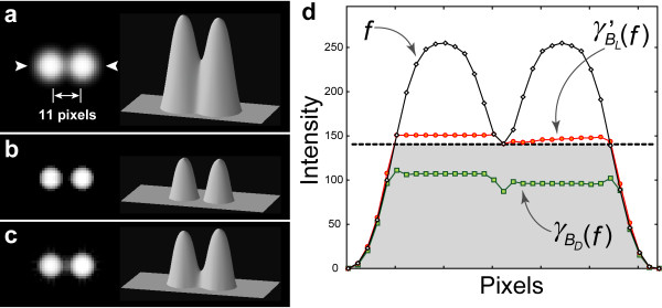
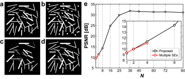
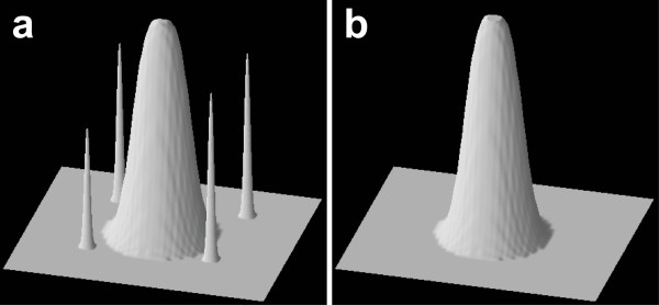
A new method to extract closely located, small target spots from biological images is proposed. This method starts with a simple but practical operation based on the extended morphological top-hat transformation to subtract an uneven background. The core of our novel approach is the following: first, the original image is rotated in an arbitrary direction and each rotated image is opened with a single straight line-segment structuring element. Second, the opened images are unified and then subtracted from the original image. To evaluate these procedures, model images of simulated spots with closely located targets were created and the efficacy of our method was compared to that of conventional morphological filtering methods. The results showed the better performance of our method. The spots of real microscope images can be quantified to confirm that the method is applicable in a given practice.

CONCLUSIONS

Our method achieved effective spot extraction under various image conditions, including aggregated target spots, poor signal-to-noise ratio, and large variations in the background intensity. Furthermore, it has no restrictions with respect to the shape of the extracted spots. The features of our method allow its broad application in biological and biomedical image information analysis.

摘要

背景

在研究细胞和组织内特定蛋白质的空间分布和动态时,需要一种可靠的提取技术来解析明场或电子显微镜图像中的多个斑点。目前,传统的图像处理方法在复杂显微镜图像中自动提取和特征提取斑点存在许多挑战。

结果

提出了一种从生物图像中提取近距离、小目标斑点的新方法。该方法首先基于扩展的形态学顶帽变换进行简单但实用的操作,以减去不均匀的背景。我们新方法的核心如下:首先,将原始图像以任意方向旋转,然后用单个直线段结构元素对每个旋转图像进行打开操作。其次,将打开的图像统一并从原始图像中减去。为了评估这些过程,创建了具有近距离目标的模拟斑点的模型图像,并将我们的方法与传统的形态滤波方法的效果进行了比较。结果表明,我们的方法具有更好的性能。可以对真实显微镜图像的斑点进行量化,以确认该方法在给定实践中的适用性。

结论

我们的方法在各种图像条件下都能有效地提取斑点,包括聚集的目标斑点、信噪比差和背景强度变化大的情况。此外,它对提取斑点的形状没有限制。我们的方法的特点使其在生物和生物医学图像信息分析中有广泛的应用。

https://cdn.ncbi.nlm.nih.gov/pmc/blobs/660d/2914730/72da1ffea8ee/1471-2105-11-373-8.jpg
https://cdn.ncbi.nlm.nih.gov/pmc/blobs/660d/2914730/6fde88e32133/1471-2105-11-373-1.jpg
https://cdn.ncbi.nlm.nih.gov/pmc/blobs/660d/2914730/eed5aa84805b/1471-2105-11-373-2.jpg
https://cdn.ncbi.nlm.nih.gov/pmc/blobs/660d/2914730/ec4d51c4dc83/1471-2105-11-373-3.jpg
https://cdn.ncbi.nlm.nih.gov/pmc/blobs/660d/2914730/2c3e12d06c2c/1471-2105-11-373-4.jpg
https://cdn.ncbi.nlm.nih.gov/pmc/blobs/660d/2914730/408480ac5817/1471-2105-11-373-5.jpg
https://cdn.ncbi.nlm.nih.gov/pmc/blobs/660d/2914730/82ee8c540d3c/1471-2105-11-373-6.jpg
https://cdn.ncbi.nlm.nih.gov/pmc/blobs/660d/2914730/24a7bcee9391/1471-2105-11-373-7.jpg
https://cdn.ncbi.nlm.nih.gov/pmc/blobs/660d/2914730/72da1ffea8ee/1471-2105-11-373-8.jpg

相似文献

1
Extended morphological processing: a practical method for automatic spot detection of biological markers from microscopic images.扩展形态学处理:一种从微观图像中自动检测生物标记物的实用方法。
BMC Bioinformatics. 2010 Jul 8;11:373. doi: 10.1186/1471-2105-11-373.
2
Mathematical algorithms for automatic search, recognition, and detection of spots in ELISPOT assay.用于在酶联免疫斑点试验(ELISPOT)中自动搜索、识别和检测斑点的数学算法。
Methods Mol Biol. 2012;792:145-53. doi: 10.1007/978-1-61779-325-7_12.
3
Mixture Modeling of 2-D Gel Electrophoresis Spots Enhances the Performance of Spot Detection.二维凝胶电泳斑点的混合建模提高了斑点检测的性能。
IEEE Trans Nanobioscience. 2017 Mar;16(2):91-99. doi: 10.1109/TNB.2017.2676725. Epub 2017 Mar 1.
4
Improving gene quantification by adjustable spot-image restoration.通过可调斑点图像恢复提高基因定量分析
Bioinformatics. 2007 Sep 1;23(17):2265-72. doi: 10.1093/bioinformatics/btm337. Epub 2007 Jun 28.
5
Fast processing of microscopic images using object-based extended depth of field.使用基于对象的扩展景深快速处理显微图像。
BMC Bioinformatics. 2016 Dec 22;17(Suppl 19):516. doi: 10.1186/s12859-016-1373-2.
6
Automatic digital ECG signal extraction and normal QRS recognition from real scene ECG images.自动从真实场景 ECG 图像中提取数字 ECG 信号和识别正常 QRS 波。
Comput Methods Programs Biomed. 2020 Apr;187:105254. doi: 10.1016/j.cmpb.2019.105254. Epub 2019 Nov 30.
7
Evaluation of three methods for retrospective correction of vignetting on medical microscopy images utilizing two open source software tools.利用两种开源软件工具评估三种用于医学显微镜图像的渐晕后向校正方法。
J Microsc. 2011 Dec;244(3):320-4. doi: 10.1111/j.1365-2818.2011.03546.x. Epub 2011 Sep 27.
8
Image analysis through feature extraction by using top-hat transform-based morphological contrast operator.通过使用基于顶帽变换的形态学对比度算子进行特征提取的图像分析。
Appl Opt. 2013 Jun 1;52(16):3777-89. doi: 10.1364/AO.52.003777.
9
Quantitative comparison and evaluation of two commercially available, two-dimensional electrophoresis image analysis software packages, Z3 and Melanie.对两款市售二维电泳图像分析软件包Z3和Melanie进行定量比较与评估。
Electrophoresis. 2002 Jul;23(14):2194-202. doi: 10.1002/1522-2683(200207)23:14<2194::AID-ELPS2194>3.0.CO;2-#.
10
Automatic analysis of DNA microarray images using mathematical morphology.使用数学形态学对DNA微阵列图像进行自动分析。
Bioinformatics. 2003 Mar 22;19(5):553-62. doi: 10.1093/bioinformatics/btg057.

引用本文的文献

1
RMPJ: An ImageJ plugin for morphological information processing in biomedical images.RMPJ:一款用于生物医学图像形态学信息处理的ImageJ插件。
PLoS Comput Biol. 2025 Apr 16;21(4):e1012992. doi: 10.1371/journal.pcbi.1012992. eCollection 2025 Apr.
2
Sandwich Enzyme-Linked Immunosorbent Assay for Quantification of Callose.用于定量检测胼胝质的夹心酶联免疫吸附测定法。
Methods Protoc. 2022 Jun 26;5(4):54. doi: 10.3390/mps5040054.
3
Signal-based optical map alignment.基于信号的光学图谱比对。

本文引用的文献

1
Automatic identification of angiogenesis in double stained images of liver tissue.自动识别肝组织双重染色图像中的血管生成。
BMC Bioinformatics. 2009 Oct 8;10 Suppl 11(Suppl 11):S13. doi: 10.1186/1471-2105-10-S11-S13.
2
Quantitative comparison of spot detection methods in fluorescence microscopy.荧光显微镜中斑点检测方法的定量比较。
IEEE Trans Med Imaging. 2010 Feb;29(2):282-301. doi: 10.1109/TMI.2009.2025127. Epub 2009 Jun 23.
3
Breast tumor xenografts: diffusion-weighted MR imaging to assess early therapy with novel apoptosis-inducing anti-DR5 antibody.
PLoS One. 2021 Sep 30;16(9):e0253102. doi: 10.1371/journal.pone.0253102. eCollection 2021.
4
Correcting anisotropic intensity in light sheet images using dehazing and image morphology.使用去雾和图像形态学校正光片图像中的各向异性强度。
APL Bioeng. 2020 Jul 1;4(3):036103. doi: 10.1063/1.5144613. eCollection 2020 Sep.
5
Irradiation-injured brain tissues can self-renew in the absence of the pivotal tumor suppressor p53 in the medaka (Oryzias latipes) embryo.在青鳉(Oryzias latipes)胚胎中,受辐射损伤的脑组织在关键肿瘤抑制因子p53缺失的情况下能够自我更新。
J Radiat Res. 2016 Jan;57(1):9-15. doi: 10.1093/jrr/rrv054. Epub 2015 Sep 25.
6
Morphological image processing for quantitative shape analysis of biomedical structures: effective contrast enhancement.形态图像处理在生物医学结构定量形状分析中的应用:有效对比度增强。
J Synchrotron Radiat. 2013 Nov;20(Pt 6):848-53. doi: 10.1107/S0909049513020761. Epub 2013 Sep 25.
7
CalloseMeasurer: a novel software solution to measure callose deposition and recognise spreading callose patterns.CalloseMeasurer:一种新型的软件解决方案,用于测量胼胝质的沉积并识别扩展的胼胝质模式。
Plant Methods. 2012 Dec 17;8(1):49. doi: 10.1186/1746-4811-8-49.
8
Gamma-ray irradiation promotes premature meiosis of spontaneously differentiating testis-ova in the testis of p53-deficient medaka (Oryzias latipes).伽马射线辐照促进了 p53 缺陷型斑马鱼(Oryzias latipes)睾丸中自发分化的睾丸-卵的过早减数分裂。
Cell Death Dis. 2012 Oct 4;3(10):e395. doi: 10.1038/cddis.2012.133.
9
Mathematical morphology-based approach to the enhancement of morphological features in medical images.基于数学形态学的医学图像形态特征增强方法。
J Clin Bioinforma. 2011 Dec 16;1:33. doi: 10.1186/2043-9113-1-33.
10
The Plant Organelles Database 2 (PODB2): an updated resource containing movie data of plant organelle dynamics.植物细胞器数据库 2(PODB2):一个更新的资源,包含植物细胞器动态的电影数据。
Plant Cell Physiol. 2011 Feb;52(2):244-53. doi: 10.1093/pcp/pcq184. Epub 2010 Nov 28.
乳腺肿瘤异种移植:采用扩散加权磁共振成像评估新型诱导凋亡抗DR5抗体的早期治疗效果
Radiology. 2008 Sep;248(3):844-51. doi: 10.1148/radiol.2483071740.
4
Bioimage informatics: a new area of engineering biology.生物图像信息学:工程生物学的一个新领域。
Bioinformatics. 2008 Sep 1;24(17):1827-36. doi: 10.1093/bioinformatics/btn346. Epub 2008 Jul 4.
5
Particle filtering for multiple object tracking in dynamic fluorescence microscopy images: application to microtubule growth analysis.动态荧光显微镜图像中多目标跟踪的粒子滤波:在微管生长分析中的应用
IEEE Trans Med Imaging. 2008 Jun;27(6):789-804. doi: 10.1109/TMI.2008.916964.
6
Over the rainbow: 25 years of confocal imaging.跨越彩虹:共聚焦成像25年
Biotechniques. 2008 Apr;44(5):643-4, 646, 648. doi: 10.2144/000112798.
7
Morphological grayscale reconstruction in image analysis: applications and efficient algorithms.图像分析中的形态学灰度重建:应用与高效算法。
IEEE Trans Image Process. 1993;2(2):176-201. doi: 10.1109/83.217222.
8
The state of the art in the analysis of two-dimensional gel electrophoresis images.二维凝胶电泳图像分析的技术现状。
Appl Microbiol Biotechnol. 2007 Oct;76(6):1223-43. doi: 10.1007/s00253-007-1128-0. Epub 2007 Aug 23.
9
Detection of molecular particles in live cells via machine learning.通过机器学习检测活细胞中的分子颗粒。
Cytometry A. 2007 Aug;71(8):563-75. doi: 10.1002/cyto.a.20404.
10
A procedure to analyze surface profiles of the protein molecules visualized by quick-freeze deep-etch replica electron microscopy.一种通过快速冷冻深度蚀刻复型电子显微镜观察来分析蛋白质分子表面轮廓的方法。
Ultramicroscopy. 2007 Jan;107(1):25-39. doi: 10.1016/j.ultramic.2006.04.012. Epub 2006 May 22.