Department of Surgery, University of Texas Medical School at Houston, Houston, TX, USA.
Exp Neurol. 2010 Oct;225(2):341-52. doi: 10.1016/j.expneurol.2010.07.005. Epub 2010 Jul 15.
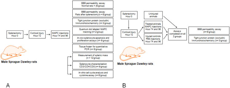
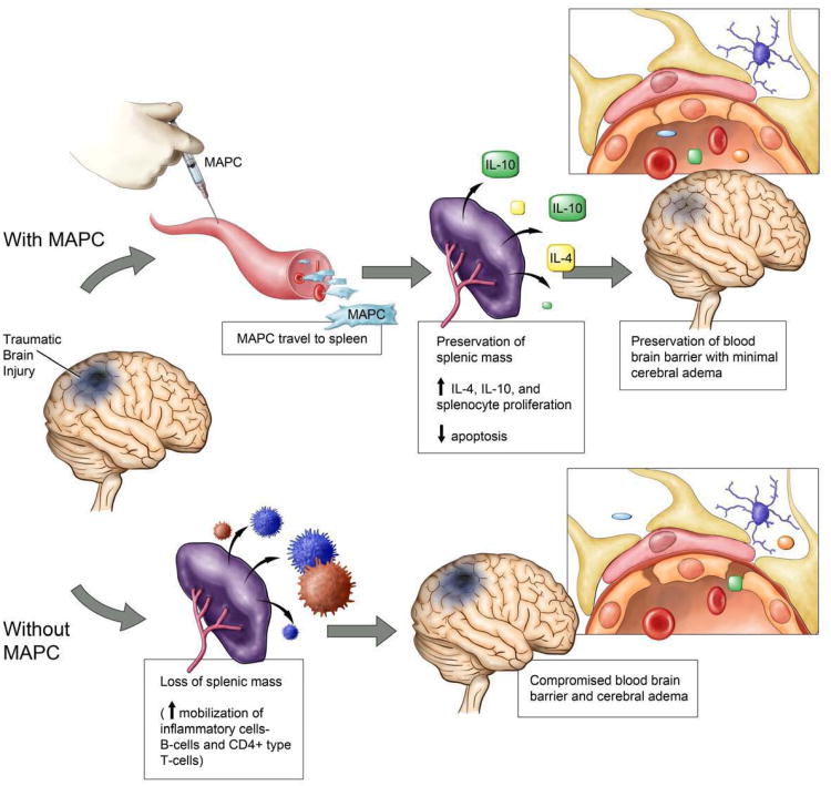
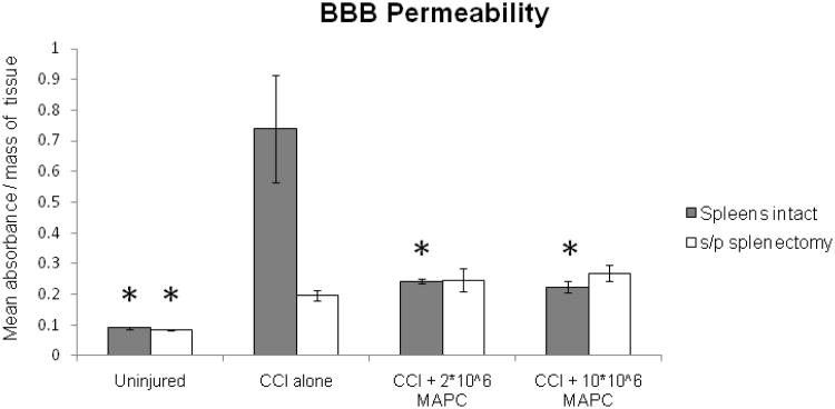
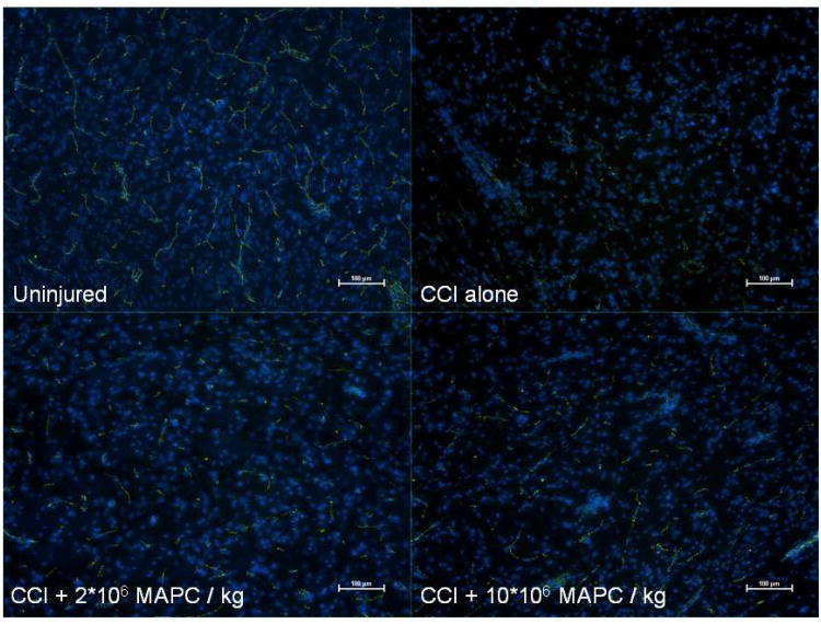
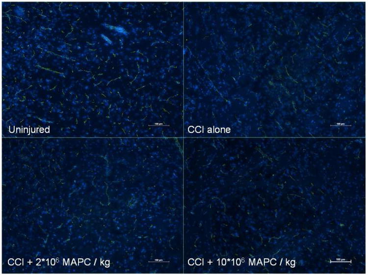
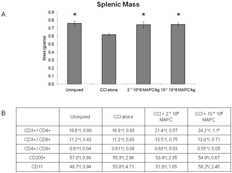
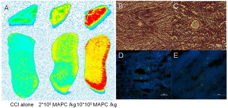
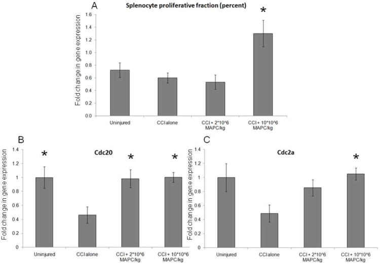
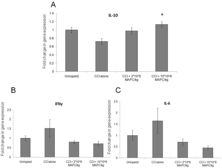
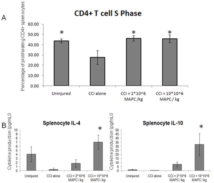
Recent investigation has shown an interaction between transplanted progenitor cells and resident splenocytes leading to the modulation of the immunologic response in neurological injury. We hypothesize that the intravenous injection of multipotent adult progenitor cells (MAPC) confers neurovascular protection after traumatic brain injury through an interaction with resident splenocytes, subsequently leading to preservation of the blood brain barrier. Four groups of rats underwent controlled cortical impact injury (3 groups) or sham injury (1 group). MAPC were injected via the tail vein at two doses (210(6) MAPC/kg or 1010(6) MAPC/kg) 2 and 24h after injury. Blood brain barrier permeability was assessed by measuring Evans blue dye extravasation (n=6/group). Additionally, splenic mass was measured (n=12/group) followed by splenocyte characterization (n=9/group) including: cell cycle analysis (n=6/group), apoptosis index (n=6/group), cell proliferation (n=6/group), and inflammatory cytokine measurements (n=6/group). Vascular architecture was determined by immunohistochemistry (n=3/group). Traumatic brain injury results in a decrease in splenic mass and increased blood brain barrier permeability. Intravenous infusion of MAPC preserved splenic mass and returned blood brain barrier permeability towards control sham injured levels. Splenocyte characterization indicated an increase in the number and proliferative rate of CD4+ T cells as well as an increase in IL-4 and IL-10 production in stimulated splenocytes isolated from the MAPC treatment groups. Immunohistochemistry demonstrated stabilization of the vascular architecture in the peri-lesion area. Traumatic brain injury causes a reduction in splenic mass that correlates with an increase in circulating immune cells leading to increased blood brain barrier permeability. The intravenous injection of MAPC preserves splenic mass and the integrity of the blood brain barrier. Furthermore, the co-localization of transplanted MAPC and resident CD4+ splenocytes is associated with a global increase in IL-4 and IL-10 production and stabilization of the cerebral microvasculature tight junction proteins.
最近的研究表明,移植的祖细胞与固有脾细胞之间存在相互作用,导致神经损伤中的免疫反应调节。我们假设,多能成体祖细胞(MAPC)静脉内注射可通过与固有脾细胞相互作用,在创伤性脑损伤后提供神经血管保护,从而导致血脑屏障的保存。四组大鼠接受皮质撞击伤(3 组)或假手术(1 组)。在损伤后 2 小时和 24 小时,通过尾静脉注射 MAPC(210(6) MAPC/kg 或 1010(6) MAPC/kg)两种剂量。通过测量 Evans 蓝染料渗出来评估血脑屏障通透性(每组 n=6)。此外,测量脾质量(每组 n=12),然后对脾细胞特征进行分析(每组 n=9),包括:细胞周期分析(每组 n=6)、凋亡指数(每组 n=6)、细胞增殖(每组 n=6)和炎症细胞因子测量(每组 n=6)。通过免疫组织化学确定血管结构(每组 n=3)。创伤性脑损伤导致脾质量下降和血脑屏障通透性增加。静脉内输注 MAPC 可保留脾质量,并使血脑屏障通透性恢复至对照假损伤水平。脾细胞特征分析表明,在 MAPC 治疗组分离的刺激脾细胞中,CD4+T 细胞的数量和增殖率增加,并且 IL-4 和 IL-10 的产生增加。免疫组织化学显示损伤周围区域血管结构的稳定。创伤性脑损伤导致脾质量减少,与循环免疫细胞增加相关,导致血脑屏障通透性增加。静脉内注射 MAPC 可保留脾质量和血脑屏障的完整性。此外,移植的 MAPC 和固有 CD4+脾细胞的共定位与 IL-4 和 IL-10 产生的整体增加以及脑微血管紧密连接蛋白的稳定相关。