University of Vienna, Faculty of Physics, Strudlhofgasse 4, A-1090 Vienna, Austria.
J Struct Biol. 2010 Dec;172(3):270-5. doi: 10.1016/j.jsb.2010.07.003. Epub 2010 Jul 15.
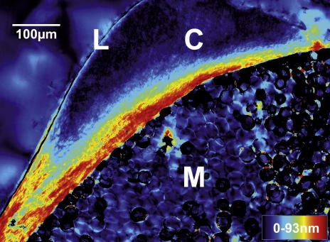
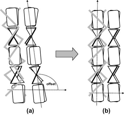
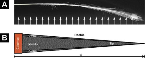
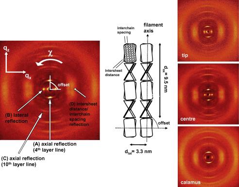
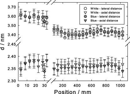
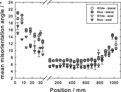
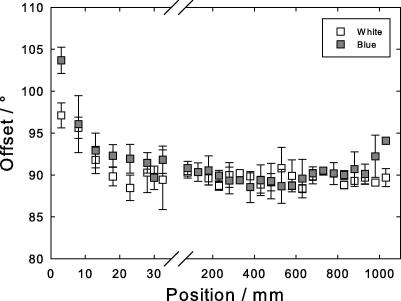
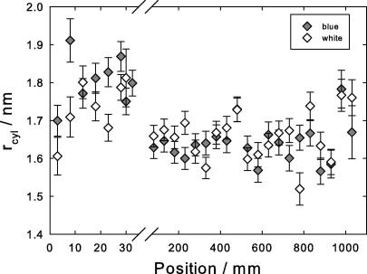
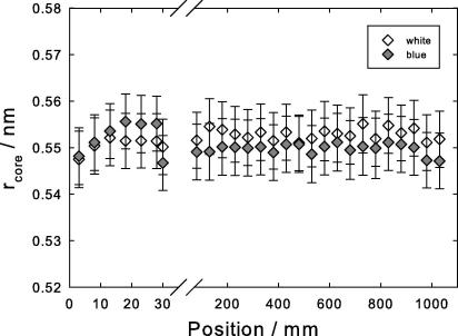
The keratin structure in the cortex of peacocks' feathers is studied by X-ray diffraction along the feather, from the calamus to the tip. It changes considerably over the first 5 cm close to the calamus and remains constant for about 1m along the length of the feather. Close to the tip, the structure loses its high degree of order. We attribute the X-ray patterns to a shrinkage of a cylindrical arrangement of β-sheets, which is not fully formed initially. In the final structure, the crystalline beta-cores are fixed by the rest of the keratin molecule. The hydrophobic residues of the beta-core are locked into a zip-like arrangement. Structurally there is no difference between the blue and the white bird.
孔雀羽毛皮质层中的角蛋白结构通过沿羽轴从羽根到羽端进行 X 射线衍射进行研究。在靠近羽根的前 5 厘米处,其变化相当大,而在羽轴长度的约 1 米处保持不变。在靠近羽端的地方,结构失去了高度有序性。我们将 X 射线图谱归因于β-折叠的圆柱形排列的收缩,该排列最初并未完全形成。在最终结构中,结晶β-核由其余角蛋白分子固定。β-核的疏水性残基被锁定在类似拉链的排列中。在结构上,蓝鸟和白鸟没有区别。