Department of Pharmaceutical Sciences, School of Pharmacy, Texas Tech University Health Sciences Center, Amarillo, TX 79106, USA.
Chem Biol Interact. 2010 Oct 6;188(1):1-14. doi: 10.1016/j.cbi.2010.05.018. Epub 2010 Jun 4.
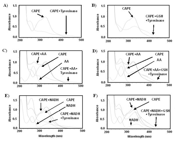
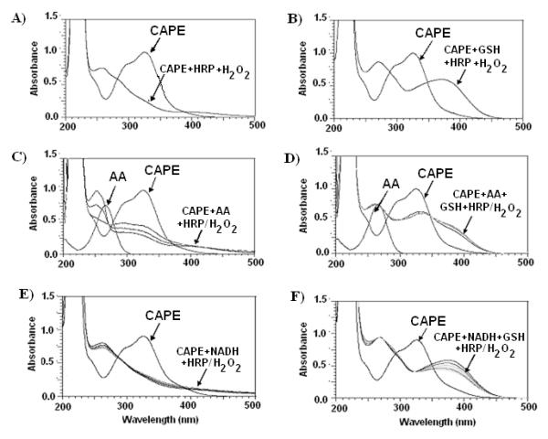
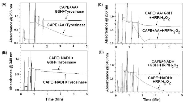
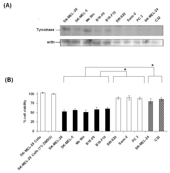
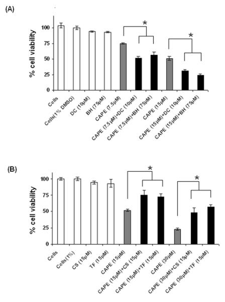
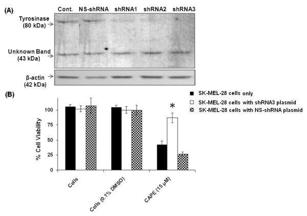
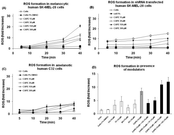
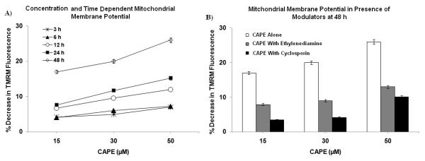
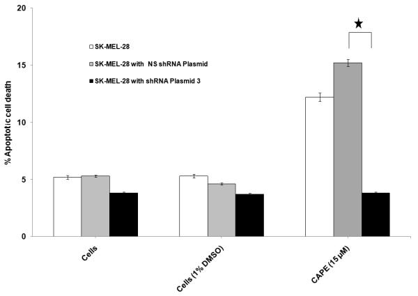
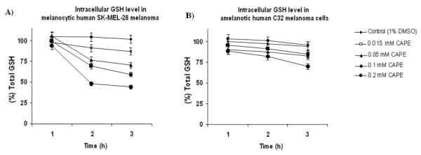
In the current work, we investigated the in vitro biochemical mechanism of Caffeic Acid Phenylethyl Ester (CAPE) toxicity and eight hydroxycinnamic/caffeic acid derivatives in vitro, using tyrosinase enzyme as a molecular target in human SK-MEL-28 melanoma cells. Enzymatic reaction models using tyrosinase/O(2) and HRP/H(2)O(2) were used to delineate the role of one- and two-electron oxidation. Ascorbic acid (AA), NADH and GSH depletion were used as markers of quinone formation and oxidative stress in CAPE induced toxicity in melanoma cells. Ethylenediamine, an o-quinone trap, prevented the formation of o-quinone and oxidations of AA and NADH mediated by tyrosinase bioactivation of CAPE. The IC(50) of CAPE towards SK-MEL-28 melanoma cells was 15muM. Dicoumarol, a diaphorase inhibitor, and 1-bromoheptane, a GSH depleting agent, increased CAPE's toxicity towards SK-MEL-28 cells indicating quinone formation played an important role in CAPE induced cell toxicity. Cyclosporin-A and trifluoperazine, inhibitors of the mitochondrial membrane permeability transition pore (PTP), prevented CAPE toxicity towards melanoma cells. We further investigated the role of tyrosinase in CAPE toxicity in the presence of a shRNA plasmid, targeting tyrosinase mRNA. Results from tyrosinase shRNA experiments showed that CAPE led to negligible anti-proliferative effect, apoptotic cell death and ROS formation in shRNA plasmid treated cells. Furthermore, it was also found that CAPE selectively caused escalation in the ROS formation and intracellular GSH (ICG) depletion in melanocytic human SK-MEL-28 cells which express functional tyrosinase. In contrast, CAPE did not lead to ROS formation and ICG depletion in amelanotic C32 melanoma cells, which do not express functional tyrosinase. These findings suggest that tyrosinase plays a major role in CAPE's selective toxicity towards melanocytic melanoma cell lines. Our findings suggest that the mechanisms of CAPE toxicity in SK-MEL-28 melanoma cells mediated by tyrosinase bioactivation of CAPE included quinone formation, ROS formation, intracellular GSH depletion and induced mitochondrial toxicity.
在目前的工作中,我们研究了咖啡酸苯乙酯(CAPE)毒性和八种羟基肉桂酸/咖啡酸衍生物在体外对酪氨酸酶酶的生物化学机制,用人黑色素瘤 SK-MEL-28 细胞作为分子靶标。使用酪氨酸酶/O(2)和 HRP/H(2)O(2)的酶促反应模型来描绘单电子和双电子氧化的作用。抗坏血酸 (AA)、NADH 和 GSH 耗竭被用作 CAPE 诱导的黑色素瘤细胞毒性中醌形成和氧化应激的标志物。乙二胺,一种邻醌捕获剂,可防止邻醌的形成以及 CAPE 被酪氨酸酶生物活化后 AA 和 NADH 的氧化。CAPE 对 SK-MEL-28 黑色素瘤细胞的 IC(50)为 15μM。二香豆素,一种脱偶联剂,1-溴庚烷,一种 GSH 耗竭剂,增加 CAPE 对 SK-MEL-28 细胞的毒性,表明醌的形成在 CAPE 诱导的细胞毒性中起重要作用。环孢菌素 A 和三氟拉嗪,线粒体膜通透性转换孔 (PTP) 的抑制剂,可防止 CAPE 对黑色素瘤细胞的毒性。我们进一步研究了在靶向酪氨酸酶 mRNA 的 shRNA 质粒存在下 CAPE 在酪氨酸酶毒性中的作用。来自 tyrosinase shRNA 实验的结果表明,CAPE 导致 shRNA 质粒处理的细胞中抗增殖作用、细胞凋亡和 ROS 形成可忽略不计。此外,还发现 CAPE 选择性地导致具有功能性酪氨酸酶的黑素细胞人 SK-MEL-28 细胞中 ROS 形成和细胞内 GSH(ICG)耗竭增加。相比之下,CAPE 不会导致不表达功能性酪氨酸酶的无黑色素 C32 黑色素瘤细胞中 ROS 形成和 ICG 耗竭。这些发现表明,酪氨酸酶在 CAPE 对黑素瘤细胞系的选择性毒性中起主要作用。我们的发现表明,CAPE 在 SK-MEL-28 黑色素瘤细胞中的毒性机制包括 CAPE 对酪氨酸酶的生物活化引起的醌形成、ROS 形成、细胞内 GSH 耗竭和诱导的线粒体毒性。