Goodyear Melinda J, Crewther Sheila G, Murphy Melanie J, Giummarra Loretta, Hazi Agnes, Junghans Barbara M, Crewther David P
School of Psychological Sciences, La Trobe University, Melbourne, Victoria, Australia.
Mol Vis. 2010 Aug 14;16:1610-9.
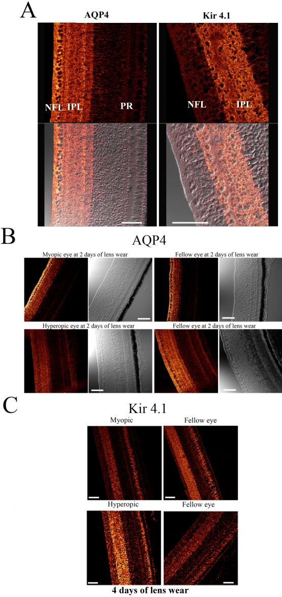
Spatial co-localization of aquaporin water channels (AQP4) and inwardly rectifying potassium ion channels (Kir4.1) on the endfeet regions of glial cells has been suggested as the basis of functionally interrelated mechanisms of osmoregulation in brain edema. The aim of this study was to investigate the spatial and temporal changes in the expression of AQP4 and Kir4.1 channels in an avascular retina during the first week of the optical induction of refractive errors.
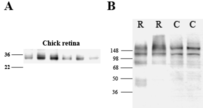
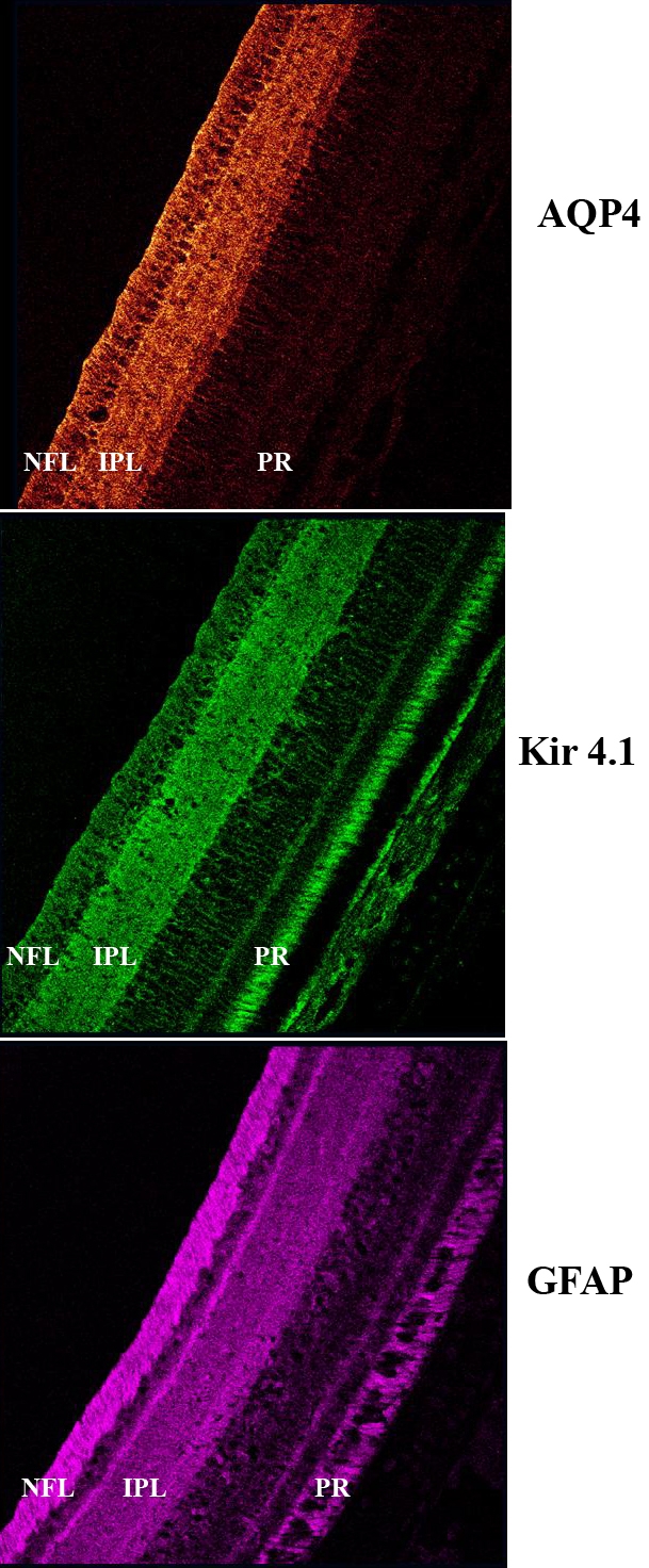
Three-day-old hatchling chicks were randomly assigned to three groups and either did not wear lenses or were monocularly goggled with +/-10D lenses for varying times up to 7 days before biometric assessment. Retinal tissue was prepared either for western blot analysis to show the presence of the AQP4 and Kir4.1 protein in the chick retina or for immunolocalization using AQP4 and Kir4.1 antibodies to determine the regional distribution and intensity of labeling during the induction of refractive errors.
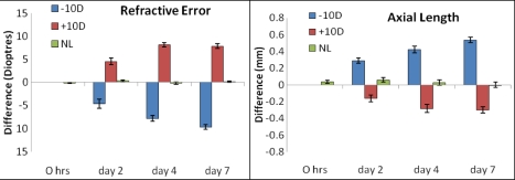
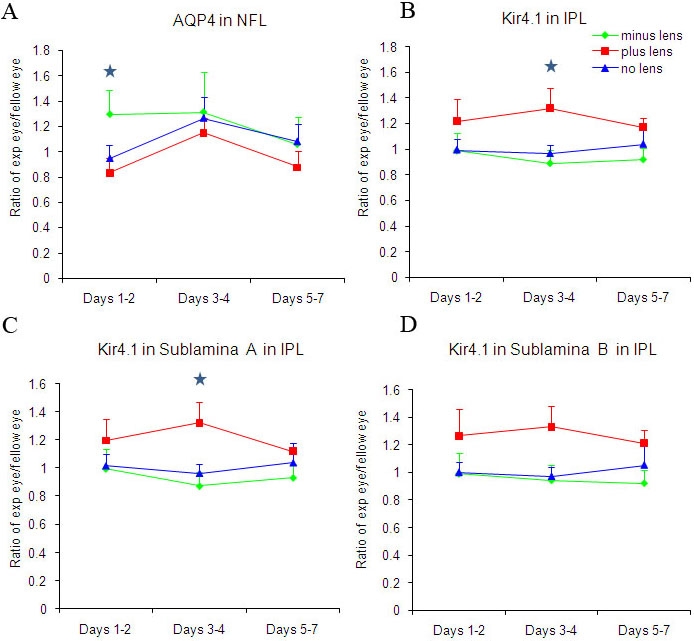
As expected, ultrasonography demonstrated that all eyes showed rapid elongation post hatching. Negative lens-wearing eyes elongated faster than fellow eyes or normal non goggled eyes and became progressively more myopic with time post lensing. Positive lens-wearing eyes showed reduced ocular growth compared to normal controls and developed a hyperopic refraction. Quantitative immunohistochemistry revealed the upregulation of AQP4 channel expression on Müller cells in the retinal nerve fiber layer during the first 2 days of negative lens wear. Kir4.1 channel upregulation in the inner plexiform layer was only found on day 4 of positive lens wear during the development of refractive hyperopia.
These results indicate that the expression of AQP4 and Kir4.1 channels on Müller cells is associated with the changes in ocular volume seen during the induction of refractive errors. However, the sites of greatest expression and the temporal pattern of the upregulation of AQP4 and Kir4.1 were dissimilar, indicating a dissociation of AQP4 and Kir4.1 function during refractive error development. Increased AQP4 expression in the nerve fiber layer is suggested to contribute to the rapid axial elongation and movement of fluid into the vitreous cavity in the presence of minus lenses; whereas, upregulation of Kir4.1 channels appears to play a role in limiting axial elongation in the presence of plus lenses.
水通道蛋白水通道(AQP4)和内向整流钾离子通道(Kir4.1)在神经胶质细胞终足区域的空间共定位被认为是脑水肿中渗透压调节功能相关机制的基础。本研究的目的是调查在屈光不正光学诱导的第一周无血管视网膜中AQP4和Kir4.1通道表达的时空变化。
将3日龄雏鸡随机分为三组,在生物测量评估前,一组不戴镜片,另外两组单眼佩戴+/-10D镜片,佩戴不同时间,最长7天。制备视网膜组织用于蛋白质印迹分析,以显示雏鸡视网膜中AQP4和Kir4.1蛋白的存在,或用于使用AQP4和Kir4.1抗体进行免疫定位,以确定屈光不正诱导期间标记的区域分布和强度。
如预期的那样,超声检查显示所有眼睛在孵化后均迅速伸长。佩戴负镜片的眼睛比未佩戴镜片的对侧眼睛或正常未佩戴眼罩的眼睛伸长更快,并且随着戴镜时间的延长逐渐变得更加近视。与正常对照组相比,佩戴正镜片的眼睛眼轴生长减缓并出现远视性屈光不正。定量免疫组织化学显示,在佩戴负镜片的前两天,视网膜神经纤维层的Müller细胞上AQP4通道表达上调。在远视性屈光不正发展过程中,仅在佩戴正镜片第4天时在内网状层发现Kir4.1通道上调。
这些结果表明,Müller细胞上AQP4和Kir4.1通道的表达与屈光不正诱导期间所见的眼容积变化有关。然而,AQP4和Kir4.1最大表达部位和上调的时间模式不同,表明在屈光不正发展过程中AQP4和Kir4.1功能解离。神经纤维层中AQP4表达增加被认为有助于在存在负镜片的情况下眼轴快速伸长以及液体进入玻璃体腔;而Kir4.1通道上调似乎在存在正镜片的情况下在限制眼轴伸长中起作用。