Goodyear Melinda J, Junghans Barbara M, Giummarra Loretta, Murphy Melanie J, Crewther David P, Crewther Sheila G
School of Psychological Science, La Trobe University, Melbourne, Australia.
Mol Vis. 2008 Feb 8;14:298-307.
Aquaporins (AQP) form a family of specialized water channels known to transport water across cell membranes and reduce osmotic gradients. The isoform AQP4 is highly expressed in the astroglia of the brain and Müller cells in the retina. In the brain, AQP4 play a role in the control of cerebral edema by shunting excess fluid into blood vessels and by upregulating during conditions of hyperosmolarity. Thus, on the basis of the hyperosmolarity seen across the retina and choroid of hatchling chickens made myopic by form deprivation (FD), we predicted an upregulation of retinal AQP4 expression during induction of myopia.
Two-day-old hatchling chicks were monocularly form-deprived for 48, 72, or 96 h, and then after biometric assessment, the eyes of these animals and the normal controls of the same age were enucleated. Retinal tissue was prepared either for western blot analysis to show the presence of the AQP4 protein in the chick retina or for immunolocalization using polyclonal AQP4 antibodies to determine regional distribution and intensity of labeling during the induction of form deprivation myopia (FDM).
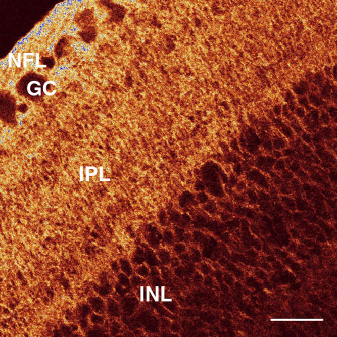
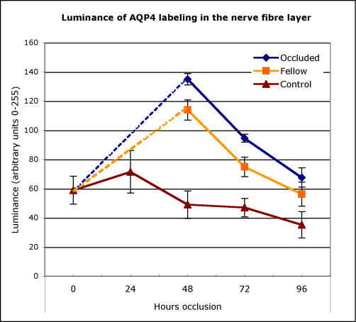
As expected, ultrasonography demonstrated that all post hatchling eyes showed rapid elongation with occluded eyes elongating faster than fellow eyes or normal controls and becoming progressively more myopic with the duration of visual deprivation. Western blot analyses revealed an approximately 30 kDa band immunoreactive for AQP4 protein and confirmed the presence of AQP4 in chicks. Immunohistochemical staining showed the greatest positive immunoreactivity for antibodies to AQP4 in the inner retina along the vitreoretinal interface, nerve fiber layer, ganglion cell layer, and inner plexiform layer in all animals. The control eyes showed relatively constant levels of AQP4 expression until day 5 after which the level appeared to decrease. By comparison, positive AQP4 immunoreactivity in the nerve fiber layer increased significantly over the first 48 h in form-deprived eyes and in fellow eyes and then decreased over the next 48 h but not to the level of expression in the normal untreated eyes.
This is the first study to demonstrate the presence of AQP4 protein in the chick retina and to associate AQP4 expression in the inner retina with the initiation of form deprivation and the period of fastest axial elongation. This increased expression of AQP4 channels near the vitread border during the time of rapid growth suggests a role for AQP4 as a conduit for movement of retinal fluid into the vitreous in form-deprived chicks.
水通道蛋白(AQP)构成了一类特殊的水通道家族,已知其可跨细胞膜转运水并降低渗透梯度。水通道蛋白4亚型(AQP4)在脑星形胶质细胞和视网膜Müller细胞中高度表达。在脑中,AQP4通过将多余液体分流至血管以及在高渗状态下上调表达,从而在控制脑水肿中发挥作用。因此,基于在形觉剥夺(FD)诱导近视的雏鸡视网膜和脉络膜中观察到的高渗现象,我们预测近视诱导过程中视网膜AQP4表达会上调。
将2日龄的雏鸡单眼形觉剥夺48、72或96小时,然后在进行生物测量评估后,摘除这些动物的眼睛以及相同年龄的正常对照的眼睛。制备视网膜组织用于蛋白质印迹分析以显示雏鸡视网膜中AQP4蛋白的存在,或用于使用多克隆AQP4抗体进行免疫定位,以确定形觉剥夺性近视(FDM)诱导过程中的区域分布和标记强度。
正如预期的那样,超声检查表明,所有孵化后的眼睛均显示出快速伸长,被遮盖眼睛的伸长速度比未遮盖眼睛或正常对照更快,并且随着视觉剥夺时间的延长,近视程度逐渐加重。蛋白质印迹分析显示出一条约30 kDa的条带,对AQP4蛋白具有免疫反应性,并证实了雏鸡中存在AQP4。免疫组织化学染色显示,在所有动物的内视网膜中,沿着玻璃体视网膜界面、神经纤维层、神经节细胞层和内网状层,AQP4抗体的阳性免疫反应最强。对照眼在第5天之前显示出相对恒定的AQP4表达水平,此后该水平似乎下降。相比之下,形觉剥夺眼和未遮盖眼的神经纤维层中AQP4阳性免疫反应性在最初48小时内显著增加,然后在接下来的48小时内下降,但未降至未处理正常眼的表达水平。
这是第一项证明雏鸡视网膜中存在AQP4蛋白,并将内视网膜中AQP4的表达与形觉剥夺的开始以及最快眼轴伸长时期相关联的研究。在快速生长期间,玻璃体边界附近AQP4通道的这种表达增加表明,AQP4在形觉剥夺雏鸡中作为视网膜液进入玻璃体的通道发挥作用。