• 文献检索
  • 文档翻译
  • 深度研究
  • 学术资讯
  • Suppr Zotero 插件Zotero 插件
  • 邀请有礼
  • 套餐&价格
  • 历史记录
应用&插件
Suppr Zotero 插件Zotero 插件浏览器插件Mac 客户端Windows 客户端微信小程序
定价
高级版会员购买积分包购买API积分包
服务
文献检索文档翻译深度研究API 文档MCP 服务
关于我们
关于 Suppr公司介绍联系我们用户协议隐私条款
关注我们

Suppr 超能文献

核心技术专利:CN118964589B侵权必究
粤ICP备2023148730 号-1Suppr @ 2026

文献检索

告别复杂PubMed语法,用中文像聊天一样搜索,搜遍4000万医学文献。AI智能推荐,让科研检索更轻松。

立即免费搜索

文件翻译

保留排版,准确专业,支持PDF/Word/PPT等文件格式,支持 12+语言互译。

免费翻译文档

深度研究

AI帮你快速写综述,25分钟生成高质量综述,智能提取关键信息,辅助科研写作。

立即免费体验

用锰增强磁共振成像评估年轻清醒自由活动小鼠中黑视蛋白对视网膜内光依赖性离子调节作用。

Light-dependant intraretinal ion regulation by melanopsin in young awake and free moving mice evaluated with manganese-enhanced MRI.

作者信息

Berkowitz Bruce A, Roberts Robin, Bissig David

机构信息

Department of Anatomy and Cell Biology, Wayne State University, Detroit, MI 48201, USA.

出版信息

Mol Vis. 2010 Aug 30;16:1776-80.

PMID:20808732
原文链接:https://pmc.ncbi.nlm.nih.gov/articles/PMC2929942/
Abstract

PURPOSE

To test the hypothesis that in young, functionally blind mice, light-dependent intraretinal ion regulation occurs via melanopsin.

METHODS

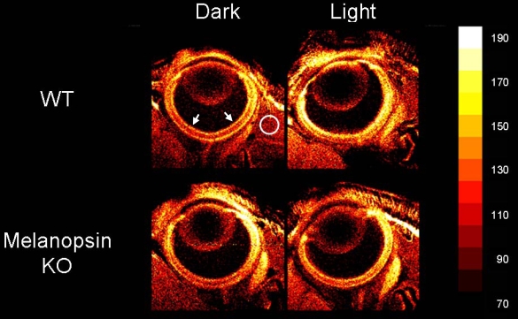
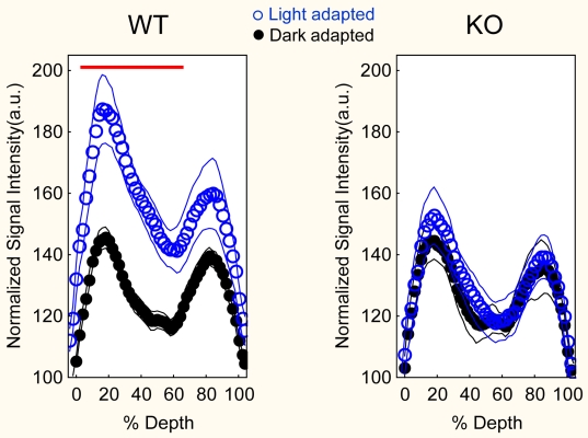
Postnatal day (P) 7 wild type (WT, C57Bl/6) and melanopsin knockout (KO, opn4-/-, B6129) mice were light or dark adapted. Awake and freely moving animals were injected intraperitoneally (ip) with MnCl(2). Four hours later, the mice in both groups were anesthetized and studied with manganese-enhanced MRI (MEMRI) to measure the extent of intraretinal uptake of manganese and whole retinal thicknesses.

RESULTS

In control P7 mice, light exposure increased (p<0.05) retinal manganese uptake over that in dark. This difference was observed throughout most of the retina. In P7 KO mice, intraretinal manganese uptake did not differ from that in age-matched dark-adapted WT mice, and was not light-dependent. No differences in whole retinal thickness were noted between groups.

CONCLUSIONS

First time evidence is presented which demonstrates intraretinal ion regulation by melanopsin in vivo.

摘要

目的

验证在年轻的功能性失明小鼠中,光依赖性视网膜内离子调节通过黑视蛋白发生这一假说。

方法

出生后第7天(P7)的野生型(WT,C57Bl/6)和黑视蛋白基因敲除(KO,opn4-/-,B6129)小鼠进行明适应或暗适应。对清醒且自由活动的动物腹腔注射(ip)氯化锰。4小时后,两组小鼠均麻醉,并采用锰增强磁共振成像(MEMRI)来测量视网膜内锰摄取程度和整个视网膜厚度。

结果

在对照P7小鼠中,光照使视网膜锰摄取量相较于黑暗环境有所增加(p<0.05)。在视网膜的大部分区域均观察到这种差异。在P7基因敲除小鼠中,视网膜内锰摄取量与年龄匹配的暗适应野生型小鼠无差异,且不依赖于光照。两组之间在整个视网膜厚度上未发现差异。

结论

首次提供了体内黑视蛋白调节视网膜内离子的证据。

https://cdn.ncbi.nlm.nih.gov/pmc/blobs/d093/2929942/a94d074013cb/mv-v16-1776-f2.jpg
https://cdn.ncbi.nlm.nih.gov/pmc/blobs/d093/2929942/5b12ab53d641/mv-v16-1776-f1.jpg
https://cdn.ncbi.nlm.nih.gov/pmc/blobs/d093/2929942/a94d074013cb/mv-v16-1776-f2.jpg
https://cdn.ncbi.nlm.nih.gov/pmc/blobs/d093/2929942/5b12ab53d641/mv-v16-1776-f1.jpg
https://cdn.ncbi.nlm.nih.gov/pmc/blobs/d093/2929942/a94d074013cb/mv-v16-1776-f2.jpg

相似文献

1
Light-dependant intraretinal ion regulation by melanopsin in young awake and free moving mice evaluated with manganese-enhanced MRI.用锰增强磁共振成像评估年轻清醒自由活动小鼠中黑视蛋白对视网膜内光依赖性离子调节作用。
Mol Vis. 2010 Aug 30;16:1776-80.
2
Melanopsin Phototransduction Contributes to Light-Evoked Choroidal Expansion and Rod L-Type Calcium Channel Function In Vivo.黑视蛋白光转导有助于体内光诱发的脉络膜扩张和视杆细胞L型钙通道功能。
Invest Ophthalmol Vis Sci. 2016 Oct 1;57(13):5314-5319. doi: 10.1167/iovs.16-20186.
3
Retinal ion regulation in a mouse model of diabetic retinopathy: natural history and the effect of Cu/Zn superoxide dismutase overexpression.糖尿病性视网膜病变小鼠模型中的视网膜离子调节:自然病程及铜/锌超氧化物歧化酶过表达的影响
Invest Ophthalmol Vis Sci. 2009 May;50(5):2351-8. doi: 10.1167/iovs.08-2918. Epub 2008 Dec 13.
4
Quantitative mapping of ion channel regulation by visual cycle activity in rodent photoreceptors in vivo.体内啮齿动物光感受器中视循环活动对离子通道调节的定量映射。
Invest Ophthalmol Vis Sci. 2009 Apr;50(4):1880-5. doi: 10.1167/iovs.08-2958. Epub 2008 Dec 5.
5
Retinal channelrhodopsin-2-mediated activity in vivo evaluated with manganese-enhanced magnetic resonance imaging.利用锰增强磁共振成像评估视网膜通道视紫红质-2在体内的活性。
Mol Vis. 2010 Jun 9;16:1059-67.
6
Genetic dissection of horizontal cell inhibitory signaling in mice in complete darkness in vivo.小鼠在完全黑暗的体内环境中水平细胞抑制信号的遗传学剖析
Invest Ophthalmol Vis Sci. 2015 May;56(5):3132-9. doi: 10.1167/iovs.15-16581.
7
Ionic dysregulatory phenotyping of pathologic retinal thinning with manganese-enhanced MRI.利用锰增强磁共振成像对病理性视网膜变薄进行离子调节表型分析。
Invest Ophthalmol Vis Sci. 2008 Jul;49(7):3178-84. doi: 10.1167/iovs.08-1720. Epub 2008 Mar 24.
8
Melanopsin as a sleep modulator: circadian gating of the direct effects of light on sleep and altered sleep homeostasis in Opn4(-/-) mice.黑视蛋白作为一种睡眠调节因子:光对睡眠直接影响的昼夜节律门控以及Opn4(-/-)小鼠睡眠稳态的改变
PLoS Biol. 2009 Jun 9;7(6):e1000125. doi: 10.1371/journal.pbio.1000125.
9
Manganese-enhanced MRI of the DBA/2J mouse model of hereditary glaucoma.遗传性青光眼DBA/2J小鼠模型的锰增强磁共振成像
Invest Ophthalmol Vis Sci. 2008 Nov;49(11):5083-8. doi: 10.1167/iovs.08-2205. Epub 2008 Jun 14.
10
Manganese-enhanced MRI studies of alterations of intraretinal ion demand in models of ocular injury.锰增强磁共振成像研究眼损伤模型中视网膜内离子需求的变化。
Invest Ophthalmol Vis Sci. 2007 Aug;48(8):3796-804. doi: 10.1167/iovs.06-1278.

引用本文的文献

1
Lifelong Changes in the Choroidal Thickness, Refractive Status, and Ocular Dimensions in C57BL/6J Mouse.C57BL/6J 小鼠脉络膜厚度、屈光状态和眼球尺寸的终身变化。
Invest Ophthalmol Vis Sci. 2024 Oct 1;65(12):26. doi: 10.1167/iovs.65.12.26.
2
Morphometric and Microstructural Changes During Murine Retinal Development Characterized Using In Vivo Optical Coherence Tomography.利用活体光学相干断层扫描技术对小鼠视网膜发育过程中的形态和微观结构变化进行研究。
Invest Ophthalmol Vis Sci. 2021 Oct 4;62(13):20. doi: 10.1167/iovs.62.13.20.
3
Applications of Manganese-Enhanced Magnetic Resonance Imaging in Ophthalmology and Visual Neuroscience.

本文引用的文献

1
Retinal channelrhodopsin-2-mediated activity in vivo evaluated with manganese-enhanced magnetic resonance imaging.利用锰增强磁共振成像评估视网膜通道视紫红质-2在体内的活性。
Mol Vis. 2010 Jun 9;16:1059-67.
2
Role of n-type voltage-dependent calcium channels in autoimmune optic neuritis.N型电压依赖性钙通道在自身免疫性视神经炎中的作用。
Ann Neurol. 2009 Jul;66(1):81-93. doi: 10.1002/ana.21668.
3
Toward clinical application of manganese-enhanced MRI of retinal function.迈向临床应用的锰增强 MRI 视网膜功能成像。
锰增强磁共振成像在眼科学和视觉神经科学中的应用。
Front Neural Circuits. 2019 May 14;13:35. doi: 10.3389/fncir.2019.00035. eCollection 2019.
4
Development of manganese-enhanced magnetic resonance imaging of the rostral ventrolateral medulla of conscious rats: Importance of normalization and comparison with other regions of interest.清醒大鼠延髓头端腹外侧区锰增强磁共振成像的发展:标准化的重要性及与其他感兴趣区域的比较
NMR Biomed. 2018 Mar;31(3). doi: 10.1002/nbm.3887. Epub 2018 Jan 12.
5
Melanopsin Phototransduction Contributes to Light-Evoked Choroidal Expansion and Rod L-Type Calcium Channel Function In Vivo.黑视蛋白光转导有助于体内光诱发的脉络膜扩张和视杆细胞L型钙通道功能。
Invest Ophthalmol Vis Sci. 2016 Oct 1;57(13):5314-5319. doi: 10.1167/iovs.16-20186.
6
MRI of Retinal Free Radical Production With Laminar Resolution In Vivo.视网膜自由基生成的体内层状分辨率磁共振成像
Invest Ophthalmol Vis Sci. 2016 Feb;57(2):577-85. doi: 10.1167/iovs.15-18972.
7
MRI of rod cell compartment-specific function in disease and treatment in vivo.体内疾病与治疗中视杆细胞区室特异性功能的磁共振成像
Prog Retin Eye Res. 2016 Mar;51:90-106. doi: 10.1016/j.preteyeres.2015.09.001. Epub 2015 Sep 4.
8
Photoreceptors in diabetic retinopathy.糖尿病视网膜病变中的光感受器
J Diabetes Investig. 2015 Jul;6(4):371-80. doi: 10.1111/jdi.12312. Epub 2015 Jan 7.
9
Genetic dissection of horizontal cell inhibitory signaling in mice in complete darkness in vivo.小鼠在完全黑暗的体内环境中水平细胞抑制信号的遗传学剖析
Invest Ophthalmol Vis Sci. 2015 May;56(5):3132-9. doi: 10.1167/iovs.15-16581.
10
Oxidative stress and light-evoked responses of the posterior segment in a mouse model of diabetic retinopathy.糖尿病视网膜病变小鼠模型中后段的氧化应激与光诱发反应
Invest Ophthalmol Vis Sci. 2015 Jan 8;56(1):606-15. doi: 10.1167/iovs.14-15687.
Brain Res Bull. 2010 Feb 15;81(2-3):333-8. doi: 10.1016/j.brainresbull.2009.06.002. Epub 2009 Jun 11.
4
Quantitative mapping of ion channel regulation by visual cycle activity in rodent photoreceptors in vivo.体内啮齿动物光感受器中视循环活动对离子通道调节的定量映射。
Invest Ophthalmol Vis Sci. 2009 Apr;50(4):1880-5. doi: 10.1167/iovs.08-2958. Epub 2008 Dec 5.
5
Intraretinal signaling by ganglion cell photoreceptors to dopaminergic amacrine neurons.神经节细胞光感受器向多巴胺能无长突细胞的视网膜内信号传导。
Proc Natl Acad Sci U S A. 2008 Sep 16;105(37):14181-6. doi: 10.1073/pnas.0803893105. Epub 2008 Sep 8.
6
The entry of manganese ions into the brain is accelerated by the activation of N-methyl-D-aspartate receptors.N-甲基-D-天冬氨酸受体的激活会加速锰离子进入大脑。
Neuroscience. 2008 Jun 23;154(2):732-40. doi: 10.1016/j.neuroscience.2008.03.080. Epub 2008 Apr 11.
7
Ionic dysregulatory phenotyping of pathologic retinal thinning with manganese-enhanced MRI.利用锰增强磁共振成像对病理性视网膜变薄进行离子调节表型分析。
Invest Ophthalmol Vis Sci. 2008 Jul;49(7):3178-84. doi: 10.1167/iovs.08-1720. Epub 2008 Mar 24.
8
Light-evoked calcium responses of isolated melanopsin-expressing retinal ganglion cells.分离出的表达黑视蛋白的视网膜神经节细胞的光诱发钙反应。
J Neurosci. 2007 Dec 5;27(49):13468-80. doi: 10.1523/JNEUROSCI.3626-07.2007.
9
High-resolution manganese-enhanced MRI of experimental retinopathy of prematurity.早产儿视网膜病变的高分辨率锰增强磁共振成像
Invest Ophthalmol Vis Sci. 2007 Oct;48(10):4733-40. doi: 10.1167/iovs.06-1516.
10
Manganese-enhanced MRI studies of alterations of intraretinal ion demand in models of ocular injury.锰增强磁共振成像研究眼损伤模型中视网膜内离子需求的变化。
Invest Ophthalmol Vis Sci. 2007 Aug;48(8):3796-804. doi: 10.1167/iovs.06-1278.