Tandon Rohit, Takkar Shibba, Kumbhkarni Shailender, Kumar Naveen, Aslam Naved, Mohan Bishav, Wander G S
Dayanand Medical College and Hospital, Unit Hero DMC Heart Institute, Tagore Nagar, Civil Lines, Ludhiana, India.
Ann Pediatr Cardiol. 2010 Jan;3(1):87-9. doi: 10.4103/0974-2069.64362.
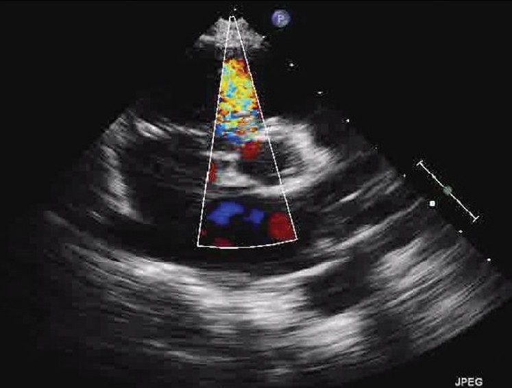
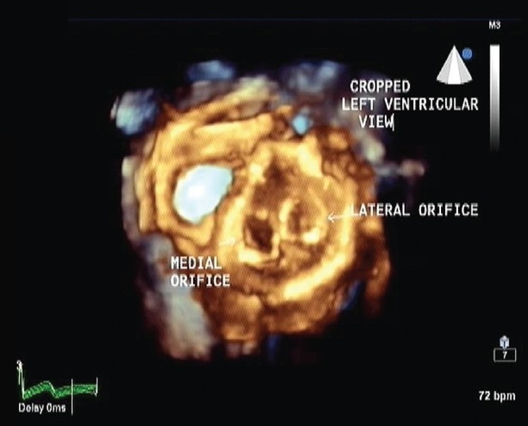
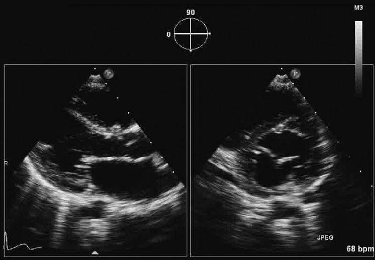
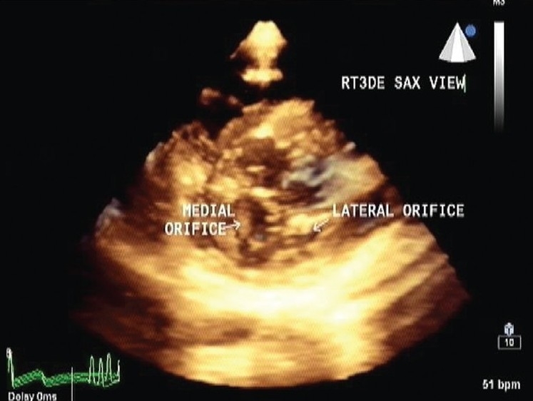
Double orifice mitral valve (DOMV) is an uncommon anomaly of surgical importance characterized by a mitral valve with a single fibrous annulus with two orifices opening into the left ventricle (LV). Subvalvular structures, especially the tensor apparatus, invariably show various degrees of abnormality. Associated congenital heart defects are common, though DOMV can occur as an isolated anomaly. Two-dimensional echocardiography is useful for diagnosis but combining it with real-time three-dimensional echocardiography helps in a more detailed evaluation of mitral valve and subvalvular structures as is shown in this case description.
双孔二尖瓣(DOMV)是一种具有手术重要性的罕见异常,其特征为二尖瓣具有单个纤维环,有两个开口通向左心室(LV)。瓣下结构,尤其是腱索装置,总是呈现出不同程度的异常。尽管DOMV可作为孤立异常出现,但相关的先天性心脏缺陷很常见。二维超声心动图对诊断有用,但将其与实时三维超声心动图相结合有助于更详细地评估二尖瓣和瓣下结构,如本病例描述所示。