• 文献检索
  • 文档翻译
  • 深度研究
  • 学术资讯
  • Suppr Zotero 插件Zotero 插件
  • 邀请有礼
  • 套餐&价格
  • 历史记录
应用&插件
Suppr Zotero 插件Zotero 插件浏览器插件Mac 客户端Windows 客户端微信小程序
定价
高级版会员购买积分包购买API积分包
服务
文献检索文档翻译深度研究API 文档MCP 服务
关于我们
关于 Suppr公司介绍联系我们用户协议隐私条款
关注我们

Suppr 超能文献

核心技术专利:CN118964589B侵权必究
粤ICP备2023148730 号-1Suppr @ 2026

文献检索

告别复杂PubMed语法,用中文像聊天一样搜索,搜遍4000万医学文献。AI智能推荐,让科研检索更轻松。

立即免费搜索

文件翻译

保留排版,准确专业,支持PDF/Word/PPT等文件格式,支持 12+语言互译。

免费翻译文档

深度研究

AI帮你快速写综述,25分钟生成高质量综述,智能提取关键信息,辅助科研写作。

立即免费体验

与变性近视相关的黄斑中心凹劈裂的病理学

Pathology of macular foveoschisis associated with degenerative myopia.

作者信息

Tang Johnny, Rivers Michael B, Moshfeghi Andrew A, Flynn Harry W, Chan Chi-Chao

机构信息

Research Service, Louis Stokes Cleveland VA Medical Center, Cleveland, OH 44106-5068, USA.

出版信息

J Ophthalmol. 2010;2010. doi: 10.1155/2010/175613. Epub 2010 Aug 12.

DOI:10.1155/2010/175613
PMID:20814547
原文链接:https://pmc.ncbi.nlm.nih.gov/articles/PMC2931386/
Abstract

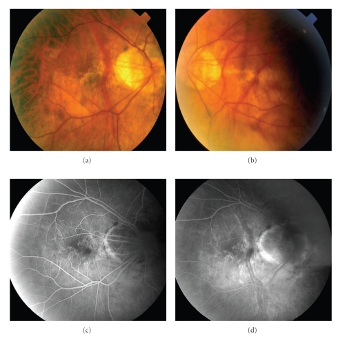
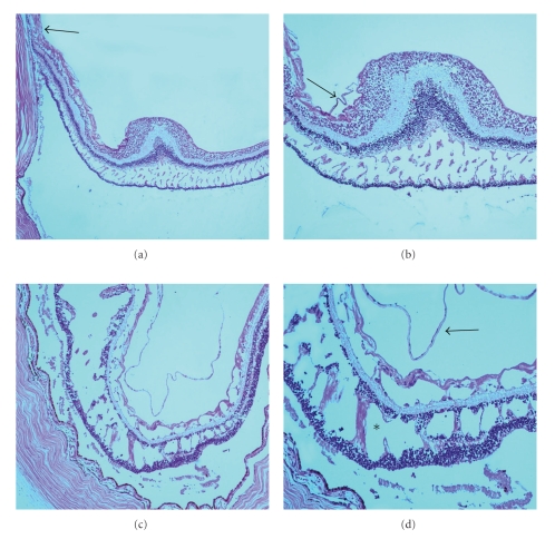
This is a clinicopathological paper on the histologic findings in myopia-associated macular foveoschisis. The findings on ophthalmic pathological study of a 73-year-old woman with high myopia are reviewed. Multiple retinoschisis cavities involving both the macula and retinal periphery were disclosed. Our paper offers tissue evidence and supports recent ocular coherence tomography reports of eyes with high myopia and associated macular foveoschisis.

摘要

这是一篇关于近视相关性黄斑中心凹劈裂组织学发现的临床病理论文。本文回顾了一名73岁高度近视女性的眼科病理研究结果。发现存在多个累及黄斑和视网膜周边部的视网膜劈裂腔。我们的论文提供了组织学证据,并支持近期关于高度近视合并黄斑中心凹劈裂眼的光学相干断层扫描报告。

https://cdn.ncbi.nlm.nih.gov/pmc/blobs/2771/2931386/fce31d144622/JOP2010-175613.002.jpg
https://cdn.ncbi.nlm.nih.gov/pmc/blobs/2771/2931386/14a3be59d645/JOP2010-175613.001.jpg
https://cdn.ncbi.nlm.nih.gov/pmc/blobs/2771/2931386/fce31d144622/JOP2010-175613.002.jpg
https://cdn.ncbi.nlm.nih.gov/pmc/blobs/2771/2931386/14a3be59d645/JOP2010-175613.001.jpg
https://cdn.ncbi.nlm.nih.gov/pmc/blobs/2771/2931386/fce31d144622/JOP2010-175613.002.jpg

相似文献

1
Pathology of macular foveoschisis associated with degenerative myopia.与变性近视相关的黄斑中心凹劈裂的病理学
J Ophthalmol. 2010;2010. doi: 10.1155/2010/175613. Epub 2010 Aug 12.
2
Relationship between macular bending and foveoschisis in myopic patients.近视患者黄斑弯曲与黄斑劈裂之间的关系。
Optom Vis Sci. 2014 May;91(5):497-506. doi: 10.1097/OPX.0000000000000250.
3
Structural profile of dome-shaped macula in degenerative myopia and its association with macular disorders.退行性近视中盘状黄斑的结构特征及其与黄斑病变的关系。
BMC Ophthalmol. 2020 May 24;20(1):202. doi: 10.1186/s12886-020-01473-2.
4
[Factors linked to foveoschisis in high myopia].[高度近视中与黄斑劈裂相关的因素]
J Fr Ophtalmol. 2014 Feb;37(2):138-42. doi: 10.1016/j.jfo.2013.11.001. Epub 2014 Jan 31.
5
Factors associated with foveoschisis and foveal detachment without macular hole in high myopia.高度近视黄斑劈裂和无黄斑裂孔的黄斑脱离的相关因素。
Eye (Lond). 2009 Feb;23(2):356-61. doi: 10.1038/sj.eye.6703038. Epub 2007 Dec 7.
6
[Vitrectomy without internal limiting membrane peeling associated with gas tamponade for treatment of foveoschisis in pathologic myopia].[玻璃体切除术联合气体填塞且不进行内界膜剥除治疗病理性近视性黄斑劈裂]
Zhonghua Yan Ke Za Zhi. 2011 Jun;47(6):497-503.
7
Visual and anatomical outcome after macular buckling for macular hole with associated foveoschisis in highly myopic eyes.高度近视黄斑裂孔伴中心凹视网膜劈裂行黄斑兜带术后的视功能和解剖学结果。
Br J Ophthalmol. 2014 Jan;98(1):104-9. doi: 10.1136/bjophthalmol-2013-304016. Epub 2013 Oct 29.
8
Evolution of full-thickness macular hole formation in a case of myopic foveoschisis.一例近视性黄斑劈裂患者全层黄斑裂孔形成的演变过程
Semin Ophthalmol. 2014 Jul;29(4):210-2. doi: 10.3109/08820538.2013.821502. Epub 2013 Sep 27.
9
Peripapillary changes detected by optical coherence tomography in eyes with high myopia.光学相干断层扫描检测高度近视眼中的视乳头周围改变。
Ophthalmology. 2007 Nov;114(11):2070-6. doi: 10.1016/j.ophtha.2007.01.016. Epub 2007 Jun 1.
10
Long-term follow-up of high myopic foveoschisis: natural course and surgical outcome.高度近视性黄斑劈裂的长期随访:自然病程与手术结果
Am J Ophthalmol. 2007 Mar;143(3):455-62. doi: 10.1016/j.ajo.2006.10.053. Epub 2006 Dec 8.

引用本文的文献

1
IMI Pathologic Myopia.病理性近视
Invest Ophthalmol Vis Sci. 2021 Apr 28;62(5):5. doi: 10.1167/iovs.62.5.5.
2
Factors Determining the Morphology of Peripapillary Retinoschisis.决定视乳头周围视网膜劈裂形态的因素
Clin Ophthalmol. 2021 Mar 25;15:1293-1300. doi: 10.2147/OPTH.S301196. eCollection 2021.
3
Outcomes of microscope-integrated intraoperative optical coherence tomography-guided center-sparing internal limiting membrane peeling for myopic traction maculopathy: a novel technique.显微镜集成术中光学相干断层扫描引导下的近视性牵引性黄斑病变中心保留内界膜剥除术的结果:一项新技术

本文引用的文献

1
Posterior vitreous detachment: evolution and complications of its early stages.后玻璃体脱离:早期演变及其并发症。
Am J Ophthalmol. 2010 Mar;149(3):371-82.e1. doi: 10.1016/j.ajo.2009.11.022.
2
Long-term follow-up of high myopic foveoschisis: natural course and surgical outcome.高度近视性黄斑劈裂的长期随访:自然病程与手术结果
Am J Ophthalmol. 2007 Mar;143(3):455-62. doi: 10.1016/j.ajo.2006.10.053. Epub 2006 Dec 8.
3
Reoperation for persistent myopic foveoschisis after primary vitrectomy.原发性玻璃体切除术后持续性近视性黄斑劈裂的再次手术
Int Ophthalmol. 2018 Aug;38(4):1689-1696. doi: 10.1007/s10792-017-0644-x. Epub 2017 Jul 4.
4
Different foveal schisis patterns in each retinal layer in eyes with hereditary juvenile retinoschisis evaluated by en-face optical coherence tomography.通过表面光学相干断层扫描评估的患有遗传性青少年视网膜劈裂症的眼睛中各视网膜层的不同黄斑劈裂模式。
Graefes Arch Clin Exp Ophthalmol. 2017 Apr;255(4):719-723. doi: 10.1007/s00417-016-3552-2. Epub 2016 Nov 16.
5
Peripapillary Retinoschisis in Glaucoma Patients.青光眼患者的视乳头周围视网膜劈裂症
J Ophthalmol. 2016;2016:1612720. doi: 10.1155/2016/1612720. Epub 2016 Mar 16.
6
Myopic foveoschisis: a clinical review.近视性黄斑劈裂:临床综述
Eye (Lond). 2015 May;29(5):593-601. doi: 10.1038/eye.2014.311. Epub 2015 Mar 6.
7
Myopic choroidal neovascularisation: current concepts and update on clinical management.近视性脉络膜新生血管:当前概念及临床管理的最新进展
Br J Ophthalmol. 2015 Mar;99(3):289-96. doi: 10.1136/bjophthalmol-2014-305131. Epub 2014 Jul 1.
8
Peripapillary retinoschisis in glaucomatous eyes.青光眼性眼中的视乳头周围视网膜劈裂症
PLoS One. 2014 Feb 28;9(2):e90129. doi: 10.1371/journal.pone.0090129. eCollection 2014.
Am J Ophthalmol. 2006 Feb;141(2):414-7. doi: 10.1016/j.ajo.2005.09.009.
4
Removal of adherent cortical vitreous plaques without removing the internal limiting membrane in the repair of macular detachments in highly myopic eyes.在高度近视眼黄斑脱离修复中,去除附着的皮质玻璃体斑块而不切除内界膜。
Retina. 2005 Apr-May;25(3):290-5. doi: 10.1097/00006982-200504000-00007.
5
Potent retinal arteriolar traction as a possible cause of myopic foveoschisis.强效视网膜小动脉牵引作为近视性黄斑劈裂的一种可能病因。
Am J Ophthalmol. 2005 Mar;139(3):462-7. doi: 10.1016/j.ajo.2004.09.078.
6
Ultrastructure of internal limiting membrane in myopic foveoschisis.近视性黄斑劈裂中内界膜的超微结构
Am J Ophthalmol. 2005 Jan;139(1):197-9. doi: 10.1016/j.ajo.2004.07.027.
7
Optical coherence tomography findings in myopic traction maculopathy.近视性牵引性黄斑病变的光学相干断层扫描结果
Arch Ophthalmol. 2004 Oct;122(10):1455-60. doi: 10.1001/archopht.122.10.1455.
8
Retinoschisis with macular retinal detachment associated with vitreomacular traction syndrome.伴有玻璃体黄斑牵引综合征的视网膜劈裂症合并黄斑视网膜脱离
Retina. 2004 Apr;24(2):307-9. doi: 10.1097/00006982-200404000-00022.
9
Vitrectomy and internal limiting membrane peeling for myopic foveoschisis.玻璃体切除术及内界膜剥除术治疗近视性黄斑劈裂
Am J Ophthalmol. 2004 Apr;137(4):719-24. doi: 10.1016/j.ajo.2003.10.019.
10
Retinal detachment at the posterior pole.后极部视网膜脱离。
Br J Ophthalmol. 1958 Dec;42(12):749-53. doi: 10.1136/bjo.42.12.749.