Kavli Institute of NanoScience, Department of BioNanoScience, TU Delft, Lorentzweg 1, 2628 CJ Delft, The Netherlands.
Nature. 2010 Sep 30;467(7315):604-7. doi: 10.1038/nature09438. Epub 2010 Sep 15.
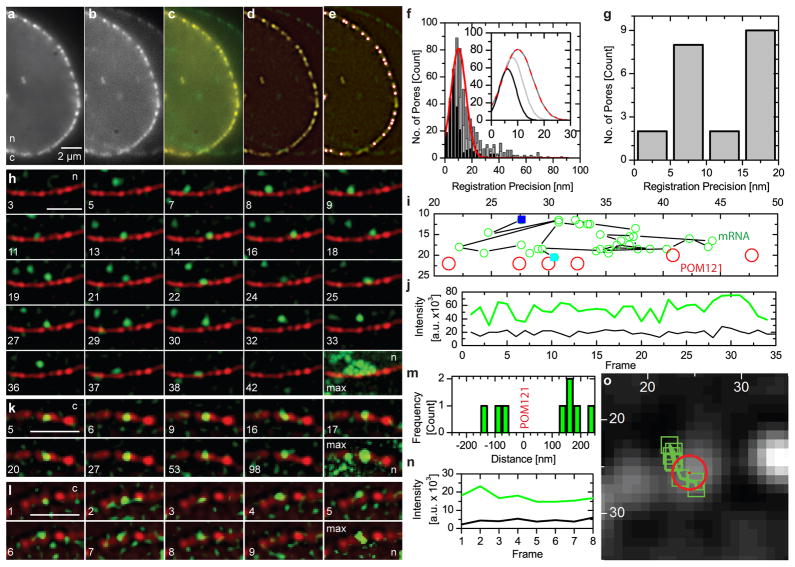
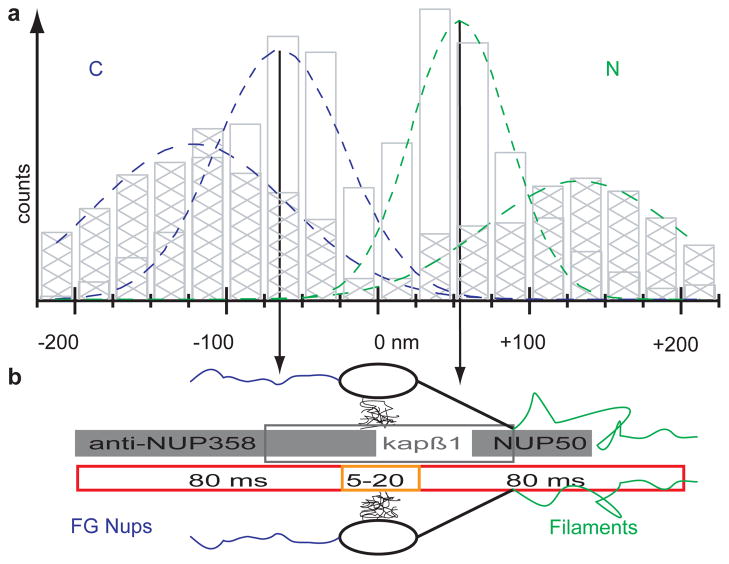
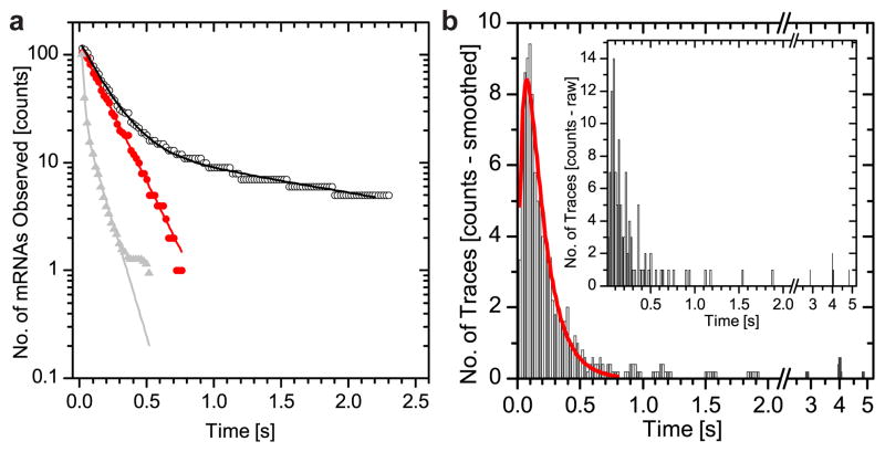
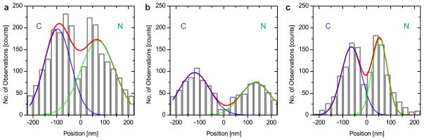
Export of messenger RNA occurs via nuclear pores, which are large nanomachines with diameters of roughly 120 nm that are the only link between the nucleus and cytoplasm. Hence, mRNA export occurs over distances smaller than the optical resolution of conventional light microscopes. There is extensive knowledge on the physical structure and composition of the nuclear pore complex, but transport selectivity and the dynamics of mRNA export at nuclear pores remain unknown. Here we developed a super-registration approach using fluorescence microscopy that can overcome the current limitations of co-localization by means of measuring intermolecular distances of chromatically different fluorescent molecules with nanometre precision. With this method we achieve 20-ms time-precision and at least 26-nm spatial precision, enabling the capture of highly transient interactions in living cells. Using this approach we were able to spatially resolve the kinetics of mRNA transport in mammalian cells and present a three-step model consisting of docking (80 ms), transport (5-20 ms) and release (80 ms), totalling 180 ± 10 ms. Notably, the translocation through the channel was not the rate-limiting step, mRNAs can move bi-directionally in the pore complex and not all pores are equally active.
信使 RNA 的输出是通过核孔进行的,核孔是一种直径约为 120nm 的大型纳米机器,是核与细胞质之间唯一的连接。因此,mRNA 的输出距离小于传统光学显微镜的分辨率。人们对核孔复合体的物理结构和组成有了广泛的了解,但 mRNA 输出的运输选择性和动力学仍然未知。在这里,我们开发了一种使用荧光显微镜的超配准方法,该方法可以通过以纳米级精度测量具有不同颜色的荧光分子之间的分子间距离,克服共定位的当前限制。通过这种方法,我们实现了 20 毫秒的时间精度和至少 26nm 的空间精度,从而能够捕获活细胞中高度瞬时的相互作用。使用这种方法,我们能够在哺乳动物细胞中空间解析 mRNA 运输的动力学,并提出了一个由对接(80ms)、运输(5-20ms)和释放(80ms)组成的三步模型,总时长为 180±10ms。值得注意的是,穿过通道的易位不是限速步骤,mRNA 可以在核孔复合体中双向移动,并非所有核孔都具有相同的活性。