• 文献检索
  • 文档翻译
  • 深度研究
  • 学术资讯
  • Suppr Zotero 插件Zotero 插件
  • 邀请有礼
  • 套餐&价格
  • 历史记录
应用&插件
Suppr Zotero 插件Zotero 插件浏览器插件Mac 客户端Windows 客户端微信小程序
定价
高级版会员购买积分包购买API积分包
服务
文献检索文档翻译深度研究API 文档MCP 服务
关于我们
关于 Suppr公司介绍联系我们用户协议隐私条款
关注我们

Suppr 超能文献

核心技术专利:CN118964589B侵权必究
粤ICP备2023148730 号-1Suppr @ 2026

文献检索

告别复杂PubMed语法,用中文像聊天一样搜索,搜遍4000万医学文献。AI智能推荐,让科研检索更轻松。

立即免费搜索

文件翻译

保留排版,准确专业,支持PDF/Word/PPT等文件格式,支持 12+语言互译。

免费翻译文档

深度研究

AI帮你快速写综述,25分钟生成高质量综述,智能提取关键信息,辅助科研写作。

立即免费体验

临床视乳头旁萎缩评估在开角型青光眼诊断中的作用。

The role of clinical parapapillary atrophy evaluation in the diagnosis of open angle glaucoma.

作者信息

Ehrlich Joshua R, Radcliffe Nathan M

机构信息

Department of Ophthalmology, Weill Cornell Medical College, New York, NY, USA.

出版信息

Clin Ophthalmol. 2010 Sep 7;4:971-6. doi: 10.2147/opth.s12420.

DOI:10.2147/opth.s12420
PMID:20856591
原文链接:https://pmc.ncbi.nlm.nih.gov/articles/PMC2938276/
Abstract

PURPOSE

To determine if clinical evaluation of parapapillary atrophy (PPA) significantly improves the ability to distinguish open-angle glaucoma (OAG) patients from glaucoma suspects.

METHODS

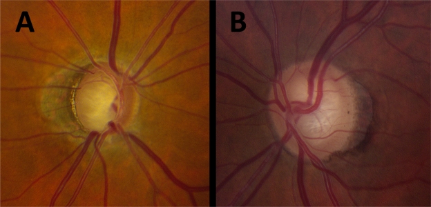
Patients in this study were under evaluation for glaucoma and had open angles, at least one reliable 24-2 SITA-standard automatic perimetry, and digital stereophotographs of the optic disc. PPA was identified clinically as a parapapillary region of absent (βPPA) or hyper/hypopigmented (αPPA) retinal pigment epithelium. A single masked observer evaluated photos for: vertical cup-to-disc ratio (CDR), clock hours of total and βPPA, βPPA as percentage width of the optic disc, presence or absence of βPPA at each disc quadrant, and ordinal rating of total PPA. Generalized linear models were used to determine odds of an abnormal or borderline glaucoma hemifield test (GHT) as a function of PPA variables and covariates; model fit was assessed using the log-likelihood ratio test.

RESULTS

Of 410 consecutive patients, 540 eyes (of 294 patients) met inclusion criteria. Mean age was greater among patients with abnormal compared with normal GHT (P < 0.001), but sex and race/ethnicity did not differ between groups (P ≥ 0.22). Age, central corneal thickness (CCT) and CDR (P ≤ 0.006), but not intraocular pressure (IOP) (P = 0.71), were significant univariable predictors of the odds of an abnormal GHT. All PPA parameters significantly predicted GHT (P ≤ 0.03), except presence of temporal βPPA (P = 0.25). Adjustment for age, CCT, IOP, and CDR reduced the association between PPA and GHT, and model fit was not greatly improved by addition of PPA variables.

CONCLUSIONS

Addition of most PPA parameters to a model already containing commonly assessed variables including age, CCT, IOP, and CDR does not significantly improve the ability to distinguish OAG patients from glaucoma suspects.

摘要

目的

确定视乳头旁萎缩(PPA)的临床评估是否能显著提高区分开角型青光眼(OAG)患者与青光眼可疑者的能力。

方法

本研究中的患者正在接受青光眼评估,具有开角、至少一次可靠的24-2 SITA标准自动视野检查以及视盘的数字立体照片。临床上将PPA识别为视网膜色素上皮缺失(βPPA)或色素沉着过度/色素沉着不足(αPPA)的视乳头旁区域。一名单盲观察者评估照片的以下指标:垂直杯盘比(CDR)、总PPA和βPPA的钟点数、βPPA占视盘宽度的百分比、每个视盘象限βPPA的有无以及总PPA的序数评分。使用广义线性模型确定异常或临界青光眼半视野检查(GHT)的几率作为PPA变量和协变量的函数;使用对数似然比检验评估模型拟合度。

结果

在410例连续患者中,540只眼(294例患者)符合纳入标准。与GHT正常的患者相比,GHT异常的患者平均年龄更大(P < 0.001),但两组之间的性别和种族/民族无差异(P≥0.22)。年龄、中央角膜厚度(CCT)和CDR(P≤0.006)是GHT异常几率的显著单变量预测因素,但眼压(IOP)不是(P = 0.71)。除颞侧βPPA的存在(P = 0.25)外,所有PPA参数均显著预测GHT(P≤0.03)。对年龄、CCT、IOP和CDR进行调整后,PPA与GHT之间的关联减弱,并且添加PPA变量后模型拟合度没有显著改善。

结论

在一个已经包含年龄、CCT、IOP和CDR等常用评估变量的模型中添加大多数PPA参数,并不会显著提高区分OAG患者与青光眼可疑者的能力。

https://cdn.ncbi.nlm.nih.gov/pmc/blobs/6106/2938276/89396a182310/opth-4-971f1.jpg

相似文献

1
The role of clinical parapapillary atrophy evaluation in the diagnosis of open angle glaucoma.临床视乳头旁萎缩评估在开角型青光眼诊断中的作用。
Clin Ophthalmol. 2010 Sep 7;4:971-6. doi: 10.2147/opth.s12420.
2
Beta-Zone parapapillary atrophy and the velocity of glaucoma progression.β 区视盘旁萎缩与青光眼进展速度。
Ophthalmology. 2010 May;117(5):909-15. doi: 10.1016/j.ophtha.2009.10.016. Epub 2010 Feb 4.
3
Association between Corneal Deformation Amplitude and Posterior Pole Profiles in Primary Open-Angle Glaucoma.原发性开角型青光眼的角膜变形幅度与后极部形态的相关性研究。
Ophthalmology. 2016 May;123(5):959-64. doi: 10.1016/j.ophtha.2015.12.043. Epub 2016 Feb 11.
4
β-Zone Parapapillary Atrophy and Rates of Glaucomatous Visual Field Progression: African Descent and Glaucoma Evaluation Study.β区视盘旁萎缩与青光眼性视野进展率:非洲裔血统与青光眼评估研究
JAMA Ophthalmol. 2017 Jun 1;135(6):617-623. doi: 10.1001/jamaophthalmol.2017.1082.
5
Anatomic relationships between disc hemorrhage and parapapillary atrophy.椎间盘出血与视乳头旁萎缩之间的解剖学关系。
Am J Ophthalmol. 2008 Nov;146(5):735-40. doi: 10.1016/j.ajo.2008.06.018. Epub 2008 Aug 23.
6
Optic Disc Change during Childhood Myopic Shift: Comparison between Eyes with an Enlarged Cup-To-Disc Ratio and Childhood Glaucoma Compared to Normal Myopic Eyes.儿童近视性屈光不正期间的视盘变化:杯盘比增大的眼睛与儿童青光眼相比,与正常近视眼睛的比较。
PLoS One. 2015 Jul 6;10(7):e0131781. doi: 10.1371/journal.pone.0131781. eCollection 2015.
7
Deep Retinal Layer Microvasculature Dropout Detected by the Optical Coherence Tomography Angiography in Glaucoma.光学相干断层扫描血管造影术检测青光眼患者视网膜深层微血管缺失
Ophthalmology. 2016 Dec;123(12):2509-2518. doi: 10.1016/j.ophtha.2016.09.002. Epub 2016 Oct 18.
8
Microstructure of β-zone parapapillary atrophy and rate of retinal nerve fiber layer thinning in primary open-angle glaucoma.原发性开角型青光眼β-区视盘旁萎缩的微观结构与视网膜神经纤维层变薄的关系。
Ophthalmology. 2014 Jul;121(7):1341-9. doi: 10.1016/j.ophtha.2014.01.008. Epub 2014 Feb 22.
9
Corneal hysteresis and Beta-zone parapapillary atrophy.角膜滞后和 Beta 区视盘旁萎缩。
Am J Ophthalmol. 2012 Feb;153(2):358-362.e1. doi: 10.1016/j.ajo.2011.06.025. Epub 2011 Sep 13.
10
Topographic correlation between β-zone parapapillary atrophy and retinal nerve fiber layer defect.β 区视盘旁萎缩与视网膜神经纤维层缺损的地形相关性。
Ophthalmology. 2013 Mar;120(3):528-534. doi: 10.1016/j.ophtha.2012.09.004. Epub 2012 Dec 1.

引用本文的文献

1
Characteristics of beta parapapillary atrophy in primary angle-closure suspect.原发性闭角型青光眼疑似患者的β 睫状视网膜神经纤维层脉络膜凹陷特征。
BMJ Open Ophthalmol. 2024 Apr 15;9(1):e001529. doi: 10.1136/bmjophth-2023-001529.
2
Peripapillary atrophy classification using CNN deep learning for glaucoma screening.基于卷积神经网络深度学习的青光眼筛查的视盘周围萎缩分类。
PLoS One. 2022 Oct 6;17(10):e0275446. doi: 10.1371/journal.pone.0275446. eCollection 2022.
3
Comparative analysis of OCT-defined parapapillary beta and gamma zones between primary open angle glaucoma and primary angle closure glaucoma.

本文引用的文献

1
Beta-Zone parapapillary atrophy and the velocity of glaucoma progression.β 区视盘旁萎缩与青光眼进展速度。
Ophthalmology. 2010 May;117(5):909-15. doi: 10.1016/j.ophtha.2009.10.016. Epub 2010 Feb 4.
2
Anatomic relationships between disc hemorrhage and parapapillary atrophy.椎间盘出血与视乳头旁萎缩之间的解剖学关系。
Am J Ophthalmol. 2008 Nov;146(5):735-40. doi: 10.1016/j.ajo.2008.06.018. Epub 2008 Aug 23.
3
Differences in parapapillary atrophy between glaucomatous and normal eyes: the Beijing Eye Study.青光眼患者与正常眼视乳头周围萎缩的差异:北京眼病研究
原发性开角型青光眼和原发性闭角型青光眼的 OCT 定义的视盘旁β区和γ区的对比分析。
Sci Rep. 2022 Jun 30;12(1):11070. doi: 10.1038/s41598-022-15457-3.
4
Controversies in the association of parapapillary atrophy with glaucoma.视乳头旁萎缩与青光眼关联中的争议。
Taiwan J Ophthalmol. 2019 Oct 31;10(4):243-249. doi: 10.4103/tjo.tjo_64_19. eCollection 2020 Oct-Dec.
5
Topographic Relationship with a Retinal Nerve Fiber Layer Defect Differs between -Zone and -Zone Parapapillary Atrophy.视盘周围萎缩α区和β区与视网膜神经纤维层缺损的地形关系有所不同。
J Ophthalmol. 2020 Aug 21;2020:6279689. doi: 10.1155/2020/6279689. eCollection 2020.
6
A novel glaucomatous representation method based on Radon and wavelet transform.基于 Radon 和小波变换的新型青光眼表现方法。
BMC Bioinformatics. 2019 Dec 24;20(Suppl 25):693. doi: 10.1186/s12859-019-3267-6.
7
Heritability of the morphology of optic nerve head and surrounding structures: The Healthy Twin Study.视神经乳头及周围结构形态的遗传力:健康双胞胎研究。
PLoS One. 2017 Nov 16;12(11):e0187498. doi: 10.1371/journal.pone.0187498. eCollection 2017.
8
Longitudinal changes in peripapillary atrophy in the ocular hypertension treatment study: a case-control assessment.眼压升高治疗研究中视乳头周围萎缩的纵向变化:病例对照评估
Ophthalmology. 2015 Jan;122(1):79-86. doi: 10.1016/j.ophtha.2014.07.033. Epub 2014 Sep 7.
Am J Ophthalmol. 2007 Oct;144(4):541-6. doi: 10.1016/j.ajo.2007.05.038. Epub 2007 Jul 25.
4
Detection and prognostic significance of optic disc hemorrhages during the Ocular Hypertension Treatment Study.眼压升高治疗研究中视盘出血的检测及其预后意义
Ophthalmology. 2006 Dec;113(12):2137-43. doi: 10.1016/j.ophtha.2006.06.022. Epub 2006 Sep 25.
5
Correlation between hemifield visual field damage and corresponding parapapillary atrophy in normal-tension glaucoma.正常眼压性青光眼中半视野视野损害与相应视乳头旁萎缩之间的相关性
Am J Ophthalmol. 2006 Jul;142(1):40-45. doi: 10.1016/j.ajo.2006.01.087.
6
Clinical implications of peripapillary atrophy in glaucoma.青光眼视乳头周围萎缩的临床意义
Curr Opin Ophthalmol. 2005 Apr;16(2):84-8. doi: 10.1097/01.icu.0000156135.20570.30.
7
Ophthalmoscopic evaluation of the parapapillary region of the optic nerve head.视神经乳头旁区域的检眼镜评估。
Klin Oczna. 2004;106(1-2 Suppl):279-89.
8
Predictive factors of the optic nerve head for development or progression of glaucomatous visual field loss.青光眼性视野缺损发生或进展的视神经乳头预测因素。
Invest Ophthalmol Vis Sci. 2004 Aug;45(8):2613-8. doi: 10.1167/iovs.03-1274.
9
Correlation between a disc hemorrhage and peripapillary atrophy in glaucoma patients with a unilateral disc hemorrhage.单侧视盘出血的青光眼患者视盘出血与视乳头周围萎缩之间的相关性
J Glaucoma. 2004 Feb;13(1):9-14. doi: 10.1097/00061198-200402000-00003.
10
Rate of optic disc cup progression in treated primary open-angle glaucoma.治疗原发性开角型青光眼时视盘杯进展率
J Glaucoma. 2003 Oct;12(5):409-16. doi: 10.1097/00061198-200310000-00003.