Department of Ophthalmology and Visual Sciences, College of Public Health, University of Nebraska Medical Center, Omaha, Nebraska, USA.
PLoS One. 2010 Aug 26;5(8):e12425. doi: 10.1371/journal.pone.0012425.
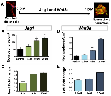
Evidence emerging from a variety of approaches used in different species suggests that Müller cell function may extend beyond its role of maintaining retinal homeostasis to that of progenitors in the adult retina. Enriched Müller cells in vitro or those that re-enter cell cycle in response to neurotoxin-damage to retina in vivo display multipotential and self-renewing capacities, the cardinal features of stem cells.
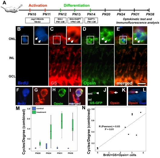
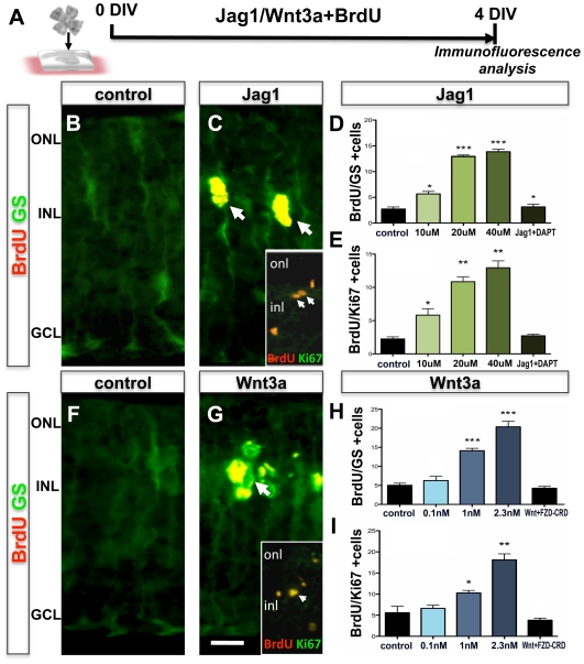
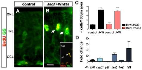
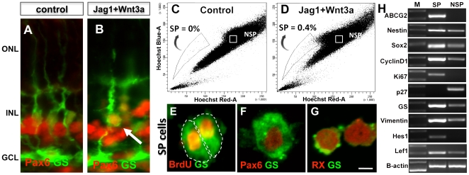
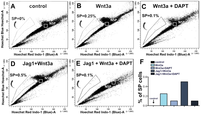
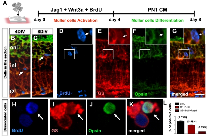
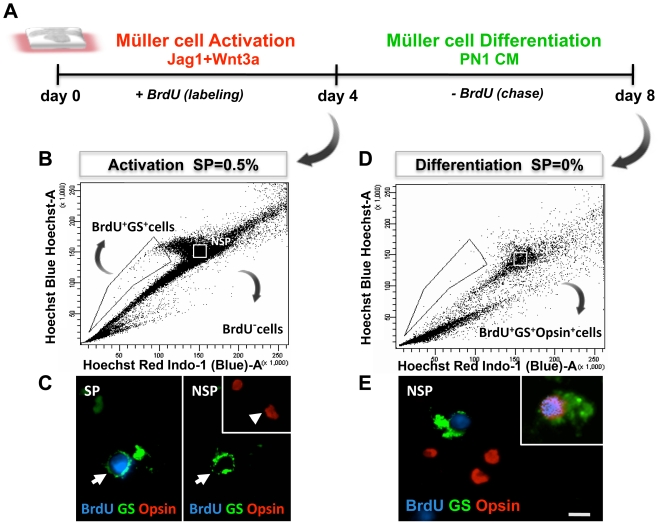
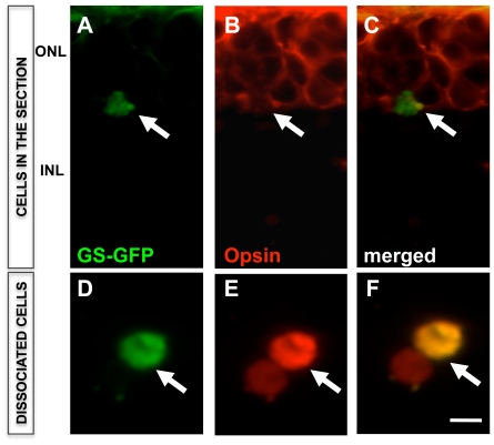
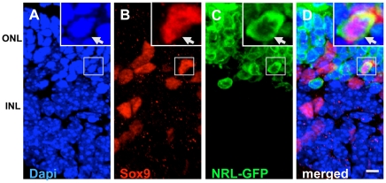
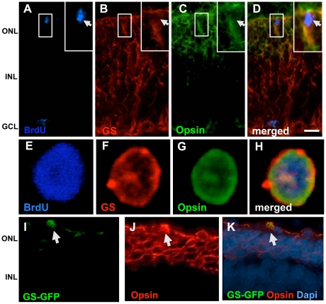
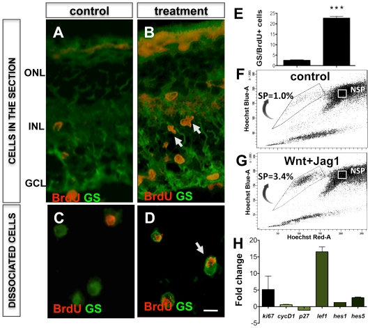
METHODOLOGY/PRINCIPAL FINDINGS: We demonstrate that Notch and Wnt signaling activate Müller cells through their canonical pathways and that a rare subset of activated Müller cells differentiates along rod photoreceptor lineage in the outer nuclear layer. The differentiation of activated Müller cells along photoreceptor lineage is confirmed by multiple approaches that included Hoechst dye efflux analysis, genetic analysis using retina from Nrl-GFP mice, and lineage tracing using GS-GFP lentivirus in wild type and rd mice in vitro and S334ter rats in vivo. Examination of S334ter rats for head-neck tracking of visual stimuli, a behavioral measure of light perception, demonstrates a significant improvement in light perception in animals treated to activate Müller cells. The number of activated Müller cells with rod photoreceptor phenotype in treated animals correlates with the improvement in their light perception.
CONCLUSION/SIGNIFICANCE: In summary, our results provide a proof of principle for non-neurotoxin-mediated activation of Müller cells through Notch and Wnt signaling toward the regeneration of rod photoreceptors.
从不同物种中使用的各种方法中涌现出的证据表明,Müller 细胞的功能可能超出了维持视网膜内稳态的作用,而成为成年视网膜中的祖细胞。体外富集的 Müller 细胞或对体内神经毒素损伤视网膜后重新进入细胞周期的 Müller 细胞表现出多能性和自我更新能力,这是干细胞的主要特征。
方法/主要发现:我们证明 Notch 和 Wnt 信号通过其经典途径激活 Müller 细胞,并且一小部分激活的 Müller 细胞在外核层中沿着杆状光感受器谱系分化。通过多种方法证实了激活的 Müller 细胞沿着光感受器谱系分化,这些方法包括 Hoechst 染料外排分析、使用 Nrl-GFP 小鼠的视网膜进行遗传分析,以及在野生型和 rd 小鼠体外和 S334ter 大鼠体内使用 GS-GFP 慢病毒进行谱系追踪。对 S334ter 大鼠进行头部-颈部跟踪视觉刺激的检查,这是一种对光感知的行为测量方法,表明用光刺激治疗可显著改善动物的光感知。经处理激活 Müller 细胞的动物中具有杆状光感受器表型的激活 Müller 细胞数量与它们的光感知改善程度相关。
结论/意义:总之,我们的结果为通过 Notch 和 Wnt 信号介导的非神经毒素激活 Müller 细胞以再生杆状光感受器提供了一个原理证明。