Biomechanics Laboratory-Rizzoli Orthopaedic Institute, Bologna, Italy.
BMC Musculoskelet Disord. 2010 Sep 27;11:220. doi: 10.1186/1471-2474-11-220.
Current research aims to develop innovative approaches to improve chondral and osteochondral regeneration. The objective of this study was to investigate the regenerative potential of platelet-rich plasma (PRP) to enhance the repair process of a collagen-hydroxyapatite scaffold in osteochondral defects in a sheep model.
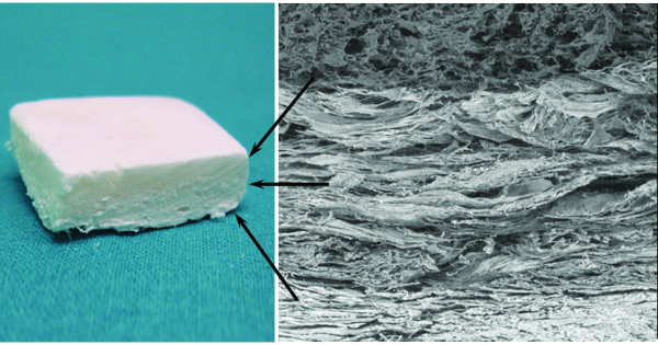
PRP was added to a new, multi-layer gradient, nanocomposite scaffold that was obtained by nucleating collagen fibrils with hydroxyapatite nanoparticles. Twenty-four osteochondral lesions were created in sheep femoral condyles. The animals were randomised to three treatment groups: scaffold, scaffold loaded with autologous PRP, and empty defect (control). The animals were sacrificed and evaluated six months after surgery.
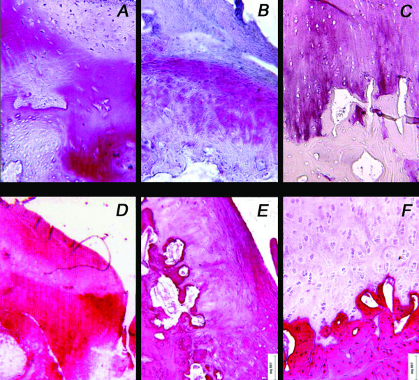
Gross evaluation and histology of the specimens showed good integration of the chondral surface in both treatment groups. Significantly better bone regeneration and cartilage surface reconstruction were observed in the group treated with the scaffold alone. Incomplete bone regeneration and irregular cartilage surface integration were observed in the group treated with the scaffold where PRP was added. In the control group, no bone and cartilage defect healing occurred; defects were filled with fibrous tissue. Quantitative macroscopic and histological score evaluations confirmed the qualitative trends observed.
The hydroxyapatite-collagen scaffold enhanced osteochondral lesion repair, but the combination with platelet growth factors did not have an additive effect; on the contrary, PRP administration had a negative effect on the results obtained by disturbing the regenerative process. In the scaffold + PRP group, highly amorphous cartilaginous repair tissue and poorly spatially organised underlying bone tissue were found.
目前的研究旨在开发创新方法来改善软骨和骨软骨再生。本研究的目的是研究富血小板血浆(PRP)在增强绵羊模型中胶原-羟磷灰石支架的软骨下骨缺损修复过程中的再生潜力。
将 PRP 添加到一种新的多层梯度纳米复合材料支架中,该支架通过将纳米羟磷灰石核晶化到胶原纤维中获得。在绵羊股骨髁上创建 24 个软骨下骨缺损。将动物随机分为三组:支架组、负载自体 PRP 的支架组和空缺陷(对照)组。术后 6 个月处死动物并进行评估。
大体评估和标本组织学检查显示两组软骨表面均有良好的整合。单独使用支架治疗的组观察到明显更好的骨再生和软骨表面重建。在添加 PRP 的支架治疗组中观察到骨再生不完全和软骨表面整合不规则。在对照组中,没有发生骨和软骨缺损愈合;缺损被纤维组织填充。宏观和组织学评分的定量评估证实了观察到的定性趋势。
羟磷灰石-胶原支架增强了软骨下骨缺损的修复,但与血小板生长因子联合使用并没有增效作用;相反,PRP 的给药通过干扰再生过程产生了负面影响。在支架+PRP 组中,发现高度非晶态软骨修复组织和空间组织不良的下骨组织。